
Page 204 - Libro vascular I
P. 204
Chap-13.qxd 1~9~04 16:43 Page 195
DUPLEX ASSESSMENT OF DEEP VENOUS THROMBOSIS AND UPPER LIMB VENOUS DISORDERS
195
GV
F
SSV
GM
SM
GM
SM
SV
A V
AB
V
A
MF
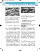
A transverse B-mode image of the posterior aspect of the mid upper calf to demonstrate the position
Figure 13.7 A: A transverse image of the common femoral vein (V) and common femoral artery (A). The common femoral vein appears distended and contains some low-level echoes. B: The common femoral vein is seen to deform but not collapse during firm transducer pressure, confirming DVT.
Figure 13.6
of the soleus muscle (SM) and a soleal vein (SV). The gastrocnemius muscle (GM) lies above the soleus muscle and is separated by a band of echogenic muscular fascia (MF). A gastrocnemius vein (GV) is seen within the muscle. The short saphenous vein (SSV) is also visible in the superficial compartment lying above the muscular fascia (F).
and sinuses located in the soleus muscle. These veins are imaged from the posterior calf (Fig. 13.6). In practice, they can be very diffi- cult to identify, especially in the normal subject.
7. Theiliacveinsareexaminedwiththepatientlying supine, as the iliac veins lie behind the bowel. The iliac veins lie slightly deeper and medial to the iliac arteries. Compression of these veins is not possible, and patency should be confirmed using color flow imaging. In addition, spectral Doppler can be used to examine flow patterns with flow augmentation maneuvers. The main limitation of examining this area is incomplete visualization due to overlying bowel gas and the potential to miss partially occluding thrombus.
8. In some cases the vena cava may need to be examined. This vessel lies to the right of the aorta when imaged in transverse section (see Fig. 11.2). Color flow imaging can be used in the transverse plane to look for filling defects, but some transverse tilt may have to be applied to the transducer to produce a reasonable Doppler angle. Flow should also be assessed in longitu- dinal section with color flow and spectral Doppler ultrasound. Examination of this area should be undertaken by a sonographer with a
considerable degree of experience. Other imag- ing modalities are generally preferable.
SCAN APPEARANCES FOR THE
ASSESSMENT OF ACUTE DVT
B-mode images
Normal appearance
The vein should appear clear and contain no echoes. In practice, there are often speckle and reverberation artifacts in the image, but the experienced sonogra- pher should have little difficulty in identifying these. Smaller veins can be difficult to distinguish from tis- sue planes. It is sometimes possible to image static or slowly moving blood as a speckle pattern within the lumen, owing to aggregation of blood cells, but the vein should collapse under transducer pressure (Figs 13.3 and 13.4). The deep calf veins can some- times be difficult to identify without the help of color flow imaging. The common femoral vein should normally distend with a Valsalva maneuver if the venous outflow through the iliac veins is patent.
Abnormal appearance
In the presence of thrombus the vein will not com- press (Fig. 13.7). In the very early stages of throm- bosis, the clot often has a degree of echogenicity due to the aggregation of red blood cells in the thrombus. Within 1 or 2 days, the clot becomes more anechoic, owing to changes occurring in the
