
Page 205 - Libro vascular I
P. 205
Chap-13.qxd 1~9~04 16:43 Page 196
196
PERIPHERAL VASCULAR ULTRASOUND
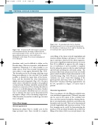
F
A transverse B-mode image of a peroneal vein thrombosis (arrow). The image is taken from the
posterolateral aspect of the calf. One trunk of the vein is grossly dilated, whereas the other is difficult to distinguish on the image. The veins are lying adjacent
to the fibula (F).
thrombus, and it can be difficult to define on the B-mode image. However, in practice, with advanced transducer technology, it is often possible to see subtle echoes. If the vein is totally occluded in the acute phase, it may appear distended (Fig. 13.8). The thrombus can be free-floating, with large areas being non-adherent to the vein wall. It is usually possible to identify the upper limit of the throm- bosis, and the thrombus tip often demonstrates slightly increased echogenicity (Fig. 13.9). The tip is much easier to identify if it extends to the popliteal or femoral veins. Prudence should be exercised with transducer compression if free-floating thrombus is present, to avoid dislodging the thrombus. Smaller areas of nonocclusive thrombus may not cause the vein to distend, but they can be demonstrated by incomplete collapse of the vein during compression. Older thrombus, beyond two weeks in age, becomes more echogenic.
Color flow images
Normal appearance
Spontaneous phasic flow is usually seen in the larger proximal veins. There should be complete
Figure 13.9 The proximal end of a free-floating thrombus (arrow) is seen in the superficial femoral vein. The thrombus is relatively anechoic and the thrombus tip is touching a valve cusp (curved arrow).
Figure 13.8
color filling of the lumen in both longitudinal and transverse planes during a calf squeeze. Color alias- ing is sometimes observed if the distal augmenta- tion causes a significant transient increase in venous flow. If it is difficult to squeeze the calf, owing to size or tenderness, it can be possible to augment flow by asking the patient to flex the ankle backward and forward, activating the calf muscle pump. The posterior tibial veins and peroneal veins are usually paired, which should be clearly demonstrated on the color flow image (Fig. 13.5). However, anatomical variations can occur. Color flow imaging of the gastrocnemius and soleal veins can be difficult, as blood flow velocities following augmentation can be low, especially if a degree of venous stasis is present.
Abnormal appearance
There is an absence of color filling in occluded veins, even with distal augmentation. Collateral veins may also be seen in the region of the occluded vein. The color flow pattern around free-floating throm- bus is very characteristic, with flow seen between the thrombus and vein wall. This can be demonstrated in both longitudinal and transverse sections. Color flow imaging can be useful for demonstrating the position of the proximal thrombus tip as full color filling of the lumen will be seen just proximal to the
