
Page 206 - Libro vascular I
P. 206
Chap-13.qxd 1~9~04 16:43 Page 197
DUPLEX ASSESSMENT OF DEEP VENOUS THROMBOSIS AND UPPER LIMB VENOUS DISORDERS
197
A
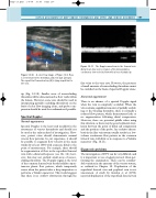
Figure 13.10 A color flow image of Figure 13.9. Flow is seen between the thrombus and vein wall (arrows). The superficial femoral artery (A) is lying superficial to the vein.
tip (Fig. 13.10). Smaller areas of nonoccluding thrombus will be demonstrated as flow voids within the lumen. However, some care should be used in interpreting partially occluding thrombosis on the basis of color flow imaging alone, and probe com- pression should be used for confirmation if possible.
Spectral Doppler
Normal appearance
Spectral Doppler is the least used modality in the assessment of venous thrombosis and should not be used as the only method of investigation. How- ever, patent veins should demonstrate normal venous flow patterns. In our experience, it should be possible to augment flow velocity in the main trunks by at least 100% with a squeeze distal to the point of measurement. For example, there should be augmentation of flow in the superficial femoral vein with a distal calf squeeze (see Ch. 12); how- ever, this may not exclude small areas of nonoc- cluding thrombus. The Doppler signal at the level of the common femoral vein should exhibit a spon- taneous phasic flow pattern, which temporarily ceases when the patient takes a deep inspiration or performs a Valsalva maneuver. This would suggest that there is no outflow obstruction through the
Figure 13.11 The Doppler waveform in the femoral vein distal to an iliac vein occlusion often demonstrates continuous low-velocity flow with a loss of phasicity.
iliac veins to the vena cava. However, the presence of small amounts of nonoccluding thrombus cannot be excluded on the basis of spectral Doppler alone.
Abnormal appearance
There is an absence of a spectral Doppler signal when the vein is completely occluded. When the vein contains a significant amount of partially occlud- ing or free-floating thrombus, there is normally a reduced flow pattern, which demonstrates little or no augmentation following distal compression. However, there are potential pitfalls when using this criterion, as there may be good collateral circu- lation between the point of distal calf compression and the position of the probe. An occlusive throm- bosis in the iliac vein system usually results in a low- volume continuous flow pattern in the common femoral vein, with little or no response to a Valsalva maneuver (Fig. 13.11).
Diagnostic problems
The investigation of DVT can be very difficult, and it is important to use a logical protocol when per- forming the examination. There can be consider- able variation in the anatomy of the venous system, as outlined in Chapter 12. Duplication of the superficial femoral vein and popliteal vein is not uncommon. A study by Gordon et al (1996) reported duplication of the superficial femoral vein
