
Page 208 - Libro vascular I
P. 208
Chap-13.qxd 1~9~04 16:43 Page 199
DUPLEX ASSESSMENT OF DEEP VENOUS THROMBOSIS AND UPPER LIMB VENOUS DISORDERS
199
as a collateral pathway in the presence of a superfi- cial femoral or popliteal vein occlusion. High-volume continuous flow recorded in the long saphenous vein should always be treated with suspicion (see Fig. 12.9).
There is considerable debate about the accuracy of duplex scanning for determining the age of thrombus, but it is generally accepted that it is possible to differentiate the acute phase, within the first week or two, from the subacute and chronic phases of venous thrombosis. However, there is much less certainty about differentiating subacute and chronic thrombus. This is due to the fact that the process of formation may not have been syn- chronous, and there are also irregularities in the process of lysis and fibrosis within the thrombus, producing a heterogeneous appearance. Many sono- graphers will not use the term ‘subacute’ in their reporting terminology because of this problem.
Recurrent thrombosis
Recurrent thrombotic events are common after acute DVT (Meissner et al 1995). There are con- siderable diagnostic problems in attempting to detect fresh thrombus in a vein that has been dam- aged by a prior DVT. If the patient has had a pre- vious scan or venogram, it is possible to check the extent of the thrombosis on the last report and compare it with the current scan. However, old reports may not be available, or the patient may not have had any previous investigations. In these situations, the vein should be examined carefully with B-mode and color flow imaging to look for areas of fresh thrombus. These will appear as ane- choic areas on the B-mode image, and color flow imaging will demonstrate filling defects. In prac- tice, this can be an extremely difficult examination to undertake. If there is a high degree of suspicion, a repeat scan can be performed a couple of days later to look for changes in the appearance of the vein or possible extension of thrombus.
OTHER PATHOLOGIC CONDITIONS THAT
CAN MIMIC DVT
There are a number of pathologic conditions that produce symptoms similar to DVT, and the sono- grapher should be able to identify these disorders.
A
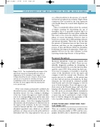
B
Figure 13.12 Two longitudinal B-mode images of the superficial femoral vein showing different stages of organization. A: The thrombus in this image is over
10 days old and has become echogenic. Areas of lysis (arrows) are seen within the thrombus. B: Partial recanalization of the vein is demonstrated with old thrombus (T), which appears fibrosed and attached
to the anterior wall.
If the vein remains permanently occluded, the thrombus becomes echogenic due to fibrosis. The thrombus retracts over time, leading to shrinkage of the vein. It may even appear as a small cord adja- cent to its corresponding artery, and in some cases the vein is difficult to differentiate from surround- ing tissue planes. Color flow imaging frequently demonstrates the development of collateral veins in the region of the occlusion. In the case of chronic common femoral and iliac vein occlusion, visible distended superficial veins, which act as collateral pathways, are often seen across the pelvis and lower abdominal wall. The long saphenous vein can act
T
