
Page 209 - Libro vascular I
P. 209
Chap-13.qxd 1~9~04 16:43 Page 200
200
PERIPHERAL VASCULAR ULTRASOUND
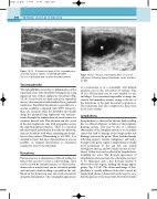
Figure 13.13 A transverse image of the long saphenous vein demonstrates evidence of thrombophlebitis.
The vein is distended and contains thrombus (arrow).
Thrombophlebitis
Thrombophlebitis occurs due to inflammation of the superficial veins, with thrombus forming in the long saphenous vein or short saphenous vein system (Fig. 13.13). It can be felt as a hard cord in the superficial tissues, often associated with localized heat, pain and tenderness. Superficial thrombosis is generally not a serious condition compared with DVT. However, there are occasions when the thrombus tip extends along the proximal long saphenous vein and pro- trudes through the saphenofemoral junction into the common femoral vein. This situation can also occur in the short saphenous vein, with propagation across the saphenopopliteal junction. There is a reported risk of proximal embolization from the thrombus tip, and care should be used when examining any throm- bus in this position (Blumenberg et al 1998). It is essential to report this type of presentation as soon as possible, as surgical intervention is sometimes required to remove the thrombus.
Hematoma
Hematomas are accumulations of blood within the tissues that can clot to form a solid swelling. They can be caused by external trauma, or other mecha- nisms such as muscle tears, can be extremely painful and can lead to limb swelling, especially in the calf. Blood in the hematoma may also track extensively along the fascial planes. The sonographic appearance
H
Figure 13.14 An area of hematoma (H) is seen in the calf muscle following injury. Hematomas can be mistaken for DVT.
of a hematoma is of a reasonably well defined anechoic area in the soft tissues or muscles (Fig. 13.14). Hematomas can be very variable in size and shape. It is sometimes impossible to image the veins in the immediate vicinity, owing to the size of the hematoma or the pain the patient experiences. The hematoma may also compress the deep veins in the local vicinity.
Lymphedema
Lymphedema is observed as chronic limb swelling due to reduced efficiency or failure of the lymphatic drainage system. This may be due to a primary abnormality of the lymphatic system or to secondary causes that lead to damage of the lymph nodes and drainage system in the groin. These include damage following surgery, trauma, malignancy and radio- therapy in the groin region. Lymphedema is usually most prominent in the calf but can extend throughout the leg, and two thirds of cases are uni- lateral. Other sites can be affected by lymphedema, including the arms. The B-mode appearance of lymphedema demonstrates the subcutaneous layer to be thickened, and a fine B-mode speckle is observed in this region, making the image appear grainy (Fig. 13.15). The ultrasound image of lym- phedema is usually different from that caused by simple fluid edema. Ultrasound can be used to con- firm the patency of the deep veins, but unfortunately
