
Page 211 - Libro vascular I
P. 211
Chap-13.qxd 1~9~04 16:44 Page 202
202
PERIPHERAL VASCULAR ULTRASOUND
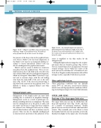
Figure 13.18
BC
V
A
A Baker’s cyst (BC) is demonstrated in this transverse image of the popliteal fossa. The popliteal
A
V
artery (A) and vein (V) are also seen in the image.
the patency of the deep veins in the popliteal fossa, even when a Baker’s cyst has been diagnosed, as the Baker’s cyst may be an incidental finding sec- ondary to venous thrombosis. Baker’s cysts can also be misdiagnosed as popliteal aneurysms.
Baker’s cysts are easiest to define in a transverse scan plane from the popliteal fossa. They are nor- mally anechoic due to the fluid in the cyst, but some may contain debris and osteocartilaginous fragments, which are echogenic. Many Baker’s cysts have a typ- ical oval or crescent shape, with the tail trailing away from the main bulk of the cyst to the joint space (Fig. 13.18). If the cyst is excessively large, it may distort the anatomy in the popliteal fossa. It is difficult to define a ruptured Baker’s cyst with ultrasound.
Enlarged lymph nodes
Enlargement of the lymph nodes can cause limb swelling due to reduction in lymphatic drainage. Enlargement occurs as a result of pathologic con- ditions, including infection or malignancy. The main sites for enlargement are at the groin or axilla, and the nodes can become so large that they compress the adjacent vein. Enlarged nodes may be tender, and localized redness and heat (erythema) may be present. They can also be clinically misdiagnosed as femoral artery aneurysms if the pulsation of the
An enlarged lymph node (arrow) is demonstrated in this transverse image at the top of
the groin. Flow is demonstrated in the lymph node. The common femoral artery (A) and vein (V) are seen below the node.
Figure 13.19
artery is amplified to the skin surface by the enlarged node.
Enlarged lymph nodes are imaged as oval or spher- ical masses that are found in groups (Fig. 13.19). They are mainly hypoechoic in appearance but may contain stronger echoes within the center of the node and can be mistaken for a thrombosed vein. Color flow Doppler usually demonstrates blood flow in larger nodes, especially if infection is present.
Other pathologic lesions
Other pathologic conditions that can clinically mimic DVT are abscesses, arteriovenous fistulas, muscle tears and hyperperfusion syndrome follow- ing arterial bypass surgery for lower limb ischemia.
UPPER LIMB VEINS
Anatomy of the deep upper limb veins
The upper limb veins can also be divided into the deep and superficial veins (Fig. 13.20), and there are a number of anatomical variations. Usually, paired veins are associated with the radial and ulnar arteries. They normally join at the elbow to form the brachial vein but can run separately to form the brachial vein higher in the upper arm. The brachial
