
Page 212 - Libro vascular I
P. 212
Chap-13.qxd 1~9~04 16:44 Page 203
DUPLEX ASSESSMENT OF DEEP VENOUS THROMBOSIS AND UPPER LIMB VENOUS DISORDERS
203
longer than the right brachiocephalic vein. It is very difficult to image the brachiocephalic veins clearly with ultrasound.
Anatomy of the superficial upper
limb veins
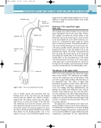
The cephalic vein and the basilic vein are the two major superficial veins in the arms (Fig. 13.20). The cephalic vein drains the dorsal surface of the hand and runs up the lateral aspect of the forearm to the antecubital fossa at the elbow and then con- tinues in a subcutaneous path along the lateral aspect of the biceps muscle. Toward the shoulder, it runs in the deltopectoral groove between the del- toid and pectoralis muscles and then pierces the clavipectoral fascia to join the axillary vein in the infraclavicular region. The basilic vein drains blood from the palm and ventral aspects of the hand and runs along the medial side of the forearm to the medial aspect of the antecubital fossa. The basilic vein then penetrates the fascia in the lower aspect of the upper arm to join the brachial vein. However, its origin can be variable, and sometimes the basilic vein may run directly into the distal axillary vein.
Thrombosis of the upper limbs
DVT is the main pathology that affects the upper limb venous system. The subclavian and axillary veins are the commonest sites for thrombosis. This can lead to upper limb swelling with distension of the superficial veins. The causes of upper limb thrombosis are similar to many of those that lead to lower limb DVT. In addition, long-term catheter access for feeding and drug administration can damage the axillary and subclavian veins. Venous thoracic outlet syndrome can also cause thrombo- sis of the subclavian vein. Effort-induced thrombosis of the subclavian vein, referred to as Paget-Schroetter syndrome, is associated with strenuous upper body exercise or repetitive movements and is often seen in younger patients.
The appearance of thrombosis in the upper extr- emities is similar to that seen in the lower extremities. A combination of compression, color flow imaging and spectral Doppler is required to confirm patency, as it is sometimes difficult to apply satisfactory probe compression, particularly in the supraclavicular
Subclavian vein
Cephalic vein
Median cubital vein
Cephalic vein
Radial vein
Figure 13.20
Internal jugular vein
Brachiocephalic vein
Axillary vein
Brachial vein Basilic vein
Ulnar vein
The venous anatomy of the arm.
vein is usually paired and associated with the brachial artery. At the top of the arm, the brachial vein becomes the axillary vein, which is usually a single trunk. The axillary vein becomes the subcla- vian vein as it crosses the border of the first rib. The subclavian vein enters the thoracic outlet but runs separately from the artery in front of the ante- rior scalene muscle. The internal jugular vein, from the neck, joins the proximal subclavian vein, which then drains via the brachiocephalic vein to the superior vena cava. The left brachiocephalic vein is
