
Page 210 - Libro vascular I
P. 210
Chap-13.qxd 1~9~04 16:43 Page 201
DUPLEX ASSESSMENT OF DEEP VENOUS THROMBOSIS AND UPPER LIMB VENOUS DISORDERS
201
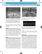
Figure 13.16 Fluid edema is demonstrated in the
subcutaneous tissues as numerous anechoic channels Figure 13.15 Lymphedema produces a grainy appearance (arrows) splaying the tissue.
in the subcutaneous tissues, as demonstrated on this transverse B-mode image. The superficial tissue is relatively thick. The muscular fascia is demonstrated by the arrow. Note the degraded image quality, typical of this disorder.
the presence of lymphedema degrades the ultrasound image, making many deep vein scans technically challenging.
Cellulitis
Cellulitis is caused by infection of the subcuta- neous tissues and skin; it produces diffuse swelling in the lower limb, often associated with pain, ten- derness and redness. There is usually evidence of edema in the region of swelling. A duplex exami- nation can confirm patency of the deep veins. In addition, there may be hyperemic flow in the veins and arteries of the limb due to the infection.
Edema
Patients can develop edema in the calf due to infec- tion, leg ulceration, local trauma, or as a result of significant venous insufficiency. This is character- ized as fluid or edema in the superficial tissues. The ultrasound appearance of edema demonstrates tis- sue splaying by numerous interstitial channels (Fig. 13.16). Patients with congestive heart failure often develop edema in the legs due to the increased pressure in the venous system and the right side of the heart. Another characteristic of congestive heart failure is the pulsatile flow pattern that is often observed in the proximal deep veins, which can be
The venous flow signals recorded from the common femoral vein of a patient with congestive
cardiac failure demonstrate a pulsatile flow pattern.
Figure 13.17
mistaken for arterial flow (Fig. 13.17). Careful attention to the color display will confirm the direction of flow.
Baker’s cysts
Baker’s cysts are bursal dilations that normally originate on the medial side of the knee between the medial head of the gastrocnemius muscle and semimembranosus tendons. A bursa is essentially a small sac of synovial fluid that prevents friction between a bone joint or tendon. The bursa can extend out of this region and into the tissue planes in the upper calf, causing swelling, pain and dis- comfort. Such bursae are caused by a number of con- ditions, including arthritis and trauma to the knee. Baker’s cysts can rupture, causing severe pain and symptoms similar to those of acute vein thrombo- sis. Large Baker’s cysts can compress the popliteal vein or deep veins of the popliteal fossa, causing a DVT. It is always necessary to identify and confirm
