
Page 203 - Libro vascular I
P. 203
Chap-13.qxd 1~9~04 16:43 Page 194
194
PERIPHERAL VASCULAR ULTRASOUND
demonstrate patency and is followed distally beyond the saphenofemoral junction, to the junc- tion of the superficial femoral vein and profunda femoris vein. The proximal segment of the pro- funda femoris vein should also be assessed for patency if possible. With the transducer turned into the longitudinal plane, the flow pattern in the common femoral vein should be assessed with color flow imaging and spectral Doppler. Flow should appear spontaneous and phasic at this level if there is no outflow obstruction. A calf squeeze can provide evidence of good flow augmentation in the proximal superficial femoral vein, which is a useful indirect indicator of probable superficial femoral and popliteal vein patency. Alternatively, strong foot flexion will also normally augment flow.
2. The superficial femoral vein is then followed in transverse section along the medial aspect of the thigh to the knee, using compression to confirm patency. The vein normally lies deep to the super- ficial femoral artery. In the adductor canal the vein may be difficult to compress. It is some- times helpful to place a hand behind the back of the lower thigh and push the flesh toward the transducer, which will bring the vein and artery more superficial to the transducer. Color flow imaging can also be used to confirm patency in this segment, but areas of nonoccluding throm- bus could be missed. Remember that duplication of the superficial femoral vein is relatively com- mon, and both trunks should be examined.
3. The popliteal vein is examined by scanning the popliteal fossa in a transverse plane. Starting in the middle of the popliteal fossa, the vein is fol- lowed proximally as far as possible to overlap the area scanned from the medial lower thigh. The popliteal vein will be seen lying above the popliteal artery when imaged from the popliteal fossa. The below-knee popliteal vein and gas- trocnemius branches are then examined in the transverse plane. The popliteal vein can also be duplicated.
4. The calf veins are often easier to identify dis- tally. They are then followed proximally to the top of the calf. The posterior tibial and peroneal veins can be imaged in a transverse plane from the medial aspect of the calf (Fig. 13.4A). From this imaging plane the peroneal veins will lie
PTV PTA
PTV
PER V PER A
PER V
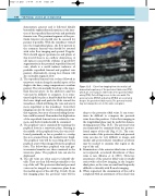
Color flow imaging from the medial calf demonstrates patency of the posterior tibial veins (PTV),
which are seen lying on either side of the posterior tibial artery (PTA). Color filling is seen to the vein walls. The peroneal veins (PER V) and artery (PER A) are seen lying deep to the posterior tibial vessels. The peroneal vessels may not always be seen in the same scan plane.
Figure 13.5
deep to the posterior tibial veins. It can some- times be difficult to compress the peroneal veins from this position. Color flow imaging in the longitudinal plane may be useful for demon- strating patency (Fig. 13.5). The peroneal veins can frequently be examined from the postero- lateral aspect of the calf (Fig. 9.11). The com- mon trunks of the posterior tibial and peroneal veins can also be very difficult to image, and medial and posterolateral transducer positions may be needed to examine this region at the top of the calf.
5. Examination of the anterior tibial veins is often not requested, as isolated thrombosis of these veins is rare (Mattos et al 1996). However, assessment of the anterior tibial veins is usually easier with color flow imaging, in the longitu- dinal plane, as the veins are small and frequently difficult to identify with B-mode imaging.
6. When requested, the examination of the calf is completed with an assessment of the soleal veins
