
Page 222 - Libro vascular I
P. 222
Chap-14.qxd 29~8~04 14:55 Page 213
GRAFT SURVEILLANCE AND PREOPERATIVE VEIN MAPPING FOR BYPASS SURGERY
Reversed vein grafts
The imaging techniques are similar to those for in situ grafts, but reversed vein grafts are frequently tunnelled deep in the thigh and, consequently, are more difficult to image. A 5MHz transducer is usually required for imaging such grafts. The graft is best located in transverse section as it divides from the native artery. The graft may drop away deeply from the proximal anastomosis. If the prox- imal anastomosis is located at the common femoral artery, the graft can be mistaken for the profunda femoris artery, or vice versa. If the graft lies deep, it may be very difficult to follow from the medial aspect of the thigh, and it can be easier to image from a posterior thigh position. If the graft is prov- ing very difficult to locate in the thigh, attempt to find a more distal segment around the level of the knee in the popliteal fossa and work upward. In extreme cases, it may be necessary to use a 3.5 MHz transducer to locate a deep segment of graft in the thigh.
Synthetic grafts
The majority of problems occurring in synthetic grafts are located at the proximal or distal anasto- mosis. It is rare for problems to develop in the main body of the graft, and a surveillance scan can often take the form of a spot check for patency combined with a more detailed assessment of the anasto- moses. It is necessary to perform a detailed assess- ment of the inflow and outflow of the graft when abnormal graft flow is recorded in the absence of any obvious graft defect.
Femoropopliteal PTFE grafts These are scanned in a similar fashion to vein grafts. The graft is often tunnelled deep in the leg.
Aortobifemoral grafts These are imaged by locating the graft at the level of the groin and following it proximally to the aorta. A combination of 5 and 3.5 MHz transducers is required for this examination.
Femorofemoral cross-over grafts These can be imaged by starting at either groin and following the graft across the pubic region to the opposite side. This can normally be achieved with a 5 MHz transducer.
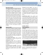
Normal B-mode image of an in situ vein graft. Note the slightly dilated area corresponding to a
valve site (arrow).
213
Iliofemoral cross-over grafts These grafts are easier to scan by starting at the distal anastomosis at the level of the femoral artery and following the graft back to the proximal anastomosis in the contralateral iliac artery. A combination of 5 and 3.5 MHz transducers is needed for this assessment. It is usually worth scanning the iliac artery above the proximal anastomosis to identify any inflow disease.
Axillobifemoral grafts These usually remain relatively superficial along their length. The cross- over section of the graft can be scanned from the distal anastomosis at the femoral artery to its bifur- cation from the main segment of the graft on the opposite side of the body. The remainder of the graft is then imaged from the ipsilateral groin, along the lateral wall of the abdomen and chest, to the infra- clavicular fossa, where the anastomosis to the axillary artery can be imaged.
B-MODE IMAGES
Normal appearance
Vein grafts
The graft lumen should be clear and of a reason- ably even caliber. Some gentle tapering is often seen in the lower portion of an in situ vein graft, as the native long saphenous vein is smaller in the lower leg. In contrast, the proximal lumen of a reversed vein graft may be smaller in caliber than the distal graft. It is common to see slight areas of dilation along a vein graft at points corresponding to valve sites (Fig. 14.7). The proximal and distal anastomoses are sometimes difficult to image clearly, due to surrounding scar tissue or depth. It may be
Figure 14.7
