
Page 223 - Libro vascular I
P. 223
Chap-14.qxd 29~8~04 14:55 Page 214
214
PERIPHERAL VASCULAR ULTRASOUND
difficult to image a deep reversed vein graft with- out color flow imaging.
Synthetic grafts
Synthetic grafts made of PTFE produce a charac- teristic image, with the anterior and posterior walls displaying a ‘double line’ appearance due to the strong reflection of ultrasound (Fig. 14.2). Some PTFE grafts are externally supported by rings that can be seen on the image (see Fig. 7.3). The cor- rugated structure of Dacron grafts, used mainly for aortobifemoral bypass surgery, is usually easy to see (see Fig. 14.16). Vein cuffs or collars are some- times used to join the graft to the distal native
Figure 14.8 In this magnified B-mode image, a large area of intimal hyperplasia (arrow) is seen in a vein graft.
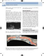
Figure 14.9 A color montage of an in situ vein graft. Areas of color flow aliasing and flow disturbance within the body of the graft may indicate a graft stenosis (arrow). These areas should be closely checked with spectral Doppler. There is retrograde filling of a short segment of the popliteal artery (curved arrow) above the distal anastomosis.
artery, and these are often seen as a short dilation at the anastomosis (Fig. 14.3).
Abnormal appearance
Many vein graft stenoses are difficult to identify with B-mode imaging alone, as they can be short or web- like and poorly echogenic. Larger areas of hyper- plasia can appear as moderately echogenic regions in the vessel lumen (Fig. 14.8). It is sometimes possible to see remnant valve cusps flapping in the lumen of in situ vein grafts due to inadequate strip- ping with the valvulotome. Areas of vein grafts may become tortuous and dilated over time, and changes in graft diameter should be recorded. In some cases large areas of thrombus or hyperplasia can be seen in aneurysmal segments, and the B-mode image may show partial stagnation or stasis of blood flow in these areas. This will be visualized as strong specular reflections in the dilated region, swirling in time with arterial pulsation. True and false aneurysms of vein or synthetic grafts can be easily seen and are discussed later in this chapter.
COLOR DOPPLER IMAGES
Normal appearance of vein grafts
An ultrasound montage of an in situ vein graft is shown in Figure 14.9. The color flow image often
