
Page 221 - Libro vascular I
P. 221
Chap-14.qxd 29~8~04 14:55 Page 212
212
PERIPHERAL VASCULAR ULTRASOUND
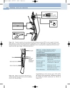
TPT Graft
B
A
CFA
Graft
Blocked SFA Profunda artery
C
C
D
Graft
SFA
CFV
Graft
Transducer positions for assessing a femoral to tibioperoneal trunk (TPT) in situ vein graft. A: Proximal graft, transverse section. B: Proximal anastomosis, longitudinal section. C: Main body of the graft, longitudinal section. D: Distal anastomosis below the popliteal fossa, longitudinal section. Scanning from a medial position below the knee may also provide a good image of the distal anastomosis.
Figure 14.5
Table 14.1 Common transducer positions for imaging the distal anastomosis of an infrainguinal graft
Level of anastomosis
Above-knee popliteal artery
Below-knee popliteal artery and tibio- peroneal trunk
Posterior tibial artery Peroneal artery
Anterior tibial artery
Transducer position
Medial aspect of lower thigh or posterior lower thigh just above popliteal fossa
Popliteal fossa or posterior and medial aspects of upper calf
Medial aspect of calf
Medial aspect of calf or from a lateral posterior position
Anterolateral aspect of calf
Fibula
Distal anastomosis
Anterior tibial artery
Vein graft
Tibia
Graft runs through interosseous membrane
Figure 14.6 Grafts to the anterior tibial artery are usually tunnelled through the interosseous membrane between the tibia and fibula.
longitudinal scar on the anterior aspect of the calf in the region of the anastomosis. Transducer positions for locating the distal anastomosis are shown in Table 14.1.
