
Page 219 - Libro vascular I
P. 219
Chap-14.qxd 29~8~04 14:55 Page 210
210
PERIPHERAL VASCULAR ULTRASOUND
require considerable commitment from the vascular laboratory, and there has been some debate as to the benefit and cost-effectiveness of surveillance pro- grams. There is, however, some evidence to suggest that they are effective in maintaining patency rates and are less costly than surgical revision after a graft thrombosis, or rehabilitation following amputation (Lundell et al 1995, Wixon et al 2000).
Synthetic grafts
The surveillance of synthetic grafts remains debat- able, as many synthetic graft occlusions occur due to spontaneous graft thrombosis. Some vascular centers perform surveillance of iliofemoral cross- over grafts and aortobifemoral grafts, particularly if there have been problems with disease in the inflow or outflow arteries. Synthetic grafts are more likely to become infected, and fluid collections or pus are sometimes found surrounding the graft at the site of infection, which frequently occurs at the groin. Graft infection is a serious complication and can cause the breakdown of the graft anastomosis, leading to uncontrollable hemorrhage. Duplex scanning has proved a useful technique for detect- ing and monitoring potential graft infections.
SYMPTOMS AND TREATMENT OF GRAFT
STENOSIS OR FAILURE
Many patients experience no symptoms in the presence of a developing graft stenosis, and grafts may fail without any prior warning. However, symptoms that can be attributable to imminent graft failure are the sudden onset of severe claudication or a sensation of coldness involving the foot. Urgent intervention is required in this situation to prevent graft occlusion. Graft surveillance pro- grams will detect the development of most graft defects, but it may be helpful to issue patients with a card providing them with information regarding their treatment and useful contact numbers should problems be suspected. Most graft stenoses are treated successfully by balloon angioplasty (see Ch. 1). However, recurrent stenoses sometimes require surgical revision involving local patching of a defect or partial graft replacement using a new segment of vein. Early graft occlusion can be treated by thrombolysis or graft thrombectomy. There is often an underlying cause for the occlusion that
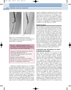
AB
Figure 14.4 A: An angiogram demonstrating a significant graft stenosis (arrow) at the distal anastomosis of a vein graft. B: The stenosis has been successfully dilated by balloon angioplasty.
Box 14.1 Suggested program for graft surveillance following discharge from hospital; time intervals are shown in months (M)
Program if no significant abnormality is detected, peak systolic velocity (PSV) ratio 2
1M, 3M, 6M, 9M,12M, program ends at 12M or continues every 6M
Program if stenosis is detected:
PSV ratio 2–2.5, reduce follow up to 2 months PSV ratio 2.6–2.9, reduce follow up to 4–6 weeks PSV ratio 3, angioplasty or graft revision
Patients are normally scanned at 1, 3, 6, 9 and 12 months following bypass surgery (Box 14.1). Many vascular units also continue to scan patients indefi- nitely beyond the first year at 6-month intervals to detect late graft problems (Erikson et al 1996). The time interval between scans is shortened to 1–2 months if a patient shows signs of developing a moderate stenosis. Patients requiring angioplasty or surgical revision of a significant graft defect recom- mence the surveillance program from the begin- ning. It can be seen that graft surveillance programs
