
Page 218 - Libro vascular I
P. 218
Chap-14.qxd 29~8~04 14:55 Page 209
GRAFT SURVEILLANCE AND PREOPERATIVE VEIN MAPPING FOR BYPASS SURGERY
anastomosis of a femoral distal graft is frequently located at the common femoral artery, although the position can vary. The position of the distal anas- tomosis is variable and depends on the distal extent of the native arterial disease. The distal anastomo- sis may lie very deep in the leg, particularly if the graft is anastomosed to the tibioperoneal trunk or peroneal artery. The natural taper of the vein along the leg matches the naturally decreasing diameter of the arteries as they run to the periphery.
In the second type of procedure, the long saphenous vein is completely removed and turned through 180° so that the distal end of the vein will form the proximal anastomosis. This is called a reversed vein graft. One particular advantage of this technique is that, in this orientation, the valves will not prevent blood flow toward the foot and do not need to be removed. Reversed vein grafts are often tunnelled deep in the thigh beneath the sartorius muscle, which can make imaging difficult. As the vein is reversed, the diameter of the proximal seg- ment of the graft is usually smaller than the distal segment. This can result in a size mismatch between the proximal inflow artery and proximal graft, which is evident on the scan. When there is insuf- ficient length of native vein available, a combina- tion of synthetic material and vein may be used to form a composite graft (Fig. 14.2).
Synthetic grafts
Synthetic grafts are used for aortobifemoral, iliofemoral, axillofemoral and femorofemoral cross- over grafts. Synthetic PTFE grafts are also used for femoropopliteal bypass, but the long-term patency
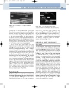
Figure 14.3
209
VEIN
Figure 14.2
vein graft.
PTFE
An example of a composite PTFE and
GRAFT
CUFF
POPLITEAL A
A vein cuff (arrow) at the distal anastomosis between a PTFE graft and popliteal artery.
rates are not as good as grafts constructed from native vein (Klinkert et al 2003). Vein cuffs or col- lars are sometimes used to join the distal end of a synthetic femoral distal graft to the native artery. They produce a localized dilation at the anastomo- sis, which is thought to reduce the risk that a stenosis will occur (Fig. 14.3).
PURPOSE OF GRAFT SURVEILLANCE
Vein grafts
The development of an intrinsic vein graft stenosis is a major source of vein graft failure. An angiogram demonstrating a graft stenosis is shown in Figure 14.4. Research has demonstrated that a significant proportion of vein grafts develop a stenosis or defect (Grigg et al 1988, Caps et al 1995). Early graft fail- ure, within the first month, is attributed to technical defects or poor patient selection. Such an example would be a patient with very poor run-off below the graft, resulting in increased resistance to flow and eventual graft thrombosis. Graft failure beyond 1 month is attributed to the development of intimal hyperplasia, which can occur when there is damage to the endothelium of the vessel wall. This causes smooth muscle proliferation into the vessel lumen and subsequent narrowing. Stenoses can occur at any point along the graft and can some- times be extremely short, web-like lesions. Incomplete removal of valve cusps during in situ bypass surgery can also cause localized flow distur- bance and narrowing. Late graft failure, beyond 12 months, can also be due to progression of athero- sclerotic disease in the native inflow or outflow arteries, above and below the graft.
