
Page 217 - Libro vascular I
P. 217
Chap-14.qxd 29~8~04 14:55 Page 208
208
PERIPHERAL VASCULAR ULTRASOUND
INTRODUCTION
Patients with significant lower limb ischemia or threatened limb loss usually require arterial bypass surgery if no other option is available to improve blood flow in the leg. Vascular surgeons are able to perform an extensive range of arterial bypass pro- cedures to restore circulation to the extremities. Bypass grafts can be made of synthetic materials, such as polytetrafluoroethylene (PTFE), or constructed from native vein, which can be assessed and marked preoperatively as described at the end of this chapter. Failure of a bypass graft due to the development of a graft stenosis is a serious complication that can result in amputation if it is not possible to unblock the graft. It is therefore common practice for vas- cular laboratories to perform regular graft surveil- lance scans to detect the development of graft defects. The majority of surveillance scans are per- formed for native vein bypass grafts below the groin (infrainguinal grafts). The surveillance of synthetic grafts is still widely practiced, but there is evidence to suggest that the benefits are less clear-cut (Lundell et al 1995). Ultrasound can also be used to image areas of potential infection following graft surgery, to see if the region of infection is in contact with the graft. The emphasis of this chapter will be on infrainguinal vein graft surveillance.
ANATOMY
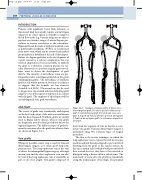
The routes of grafts vary considerably and depend on the level and extent of the native arterial disease that has been bypassed. Synthetic grafts are mainly used to bypass inflow disease, whereas vein grafts are frequently used for distal procedures below the inguinal ligament. The different types of graft fre- quently encountered in the graft surveillance clinic are shown in Figure 14.1.
Vein grafts
Whenever possible, native vein is used for femoral distal bypass surgery, as it offers good long-term patency rates. The long saphenous vein is the vein of choice for infrainguinal bypass surgery, although an arm vein and the short saphenous vein can also be used if the long saphenous vein is unsuitable in part or all of its length. Vein grafts composed of
C
A
D
F
B
E
Examples of bypass grafts. A: Above-knee femoropopliteal graft. B: Femoro-posterior tibial artery graft. C: Aortobifemoral graft. D: Iliofemoral cross-over
Figure 14.1
graft. E: Superficial femoral artery to peroneal artery graft. F: Popliteal artery bypass graft for a thrombosed popliteal aneurysm.
more than one segment of vein are known as com- posite vein grafts. Femoral distal bypass surgery is performed using two common types of surgical procedure.
The first is the in situ technique, in which the long saphenous vein is exposed but left in its native position and side branches ligated to prevent blood shunting from the graft to the venous system. As the vein contains valves which would prevent blood flow toward the foot, they have to be removed or disrupted using a device called a valvulotome. The main body of an in situ vein graft lies superficially along the medial aspect of the thigh. The proximal
