
Page 225 - Libro vascular I
P. 225
Chap-14.qxd 29~8~04 14:55 Page 216
216
PERIPHERAL VASCULAR ULTRASOUND
AB
A
B
normally assumes a pulsatile flow pattern.
large-diameter vein graft joined to a smaller out- flow artery, producing a natural velocity increase. In this situation, it is possible to see a significant increase in the PSV in the absence of a stenosis (Fig. 14.11). However, flow velocities just below the distal anastomosis should be similar to those several centimeters downstream, provided that the vessel diameter is the same. A significant stenosis would be indicated if the velocities at the anasto- mosis were found to be substantially higher (i.e., 3–4 times) than distal velocities. It is also impor- tant to ensure that the spectral Doppler angle is set correctly at the distal anastomosis, as flow is not always parallel to the vessel walls, and this can lead to errors in velocity measurements.
Abnormal appearance
Graft stenoses are categorized using a similar method to that for grading lower limb arterial disease. The PSV across the stenosis is divided by the PSV in a normal segment of graft just proximal to the stenosis (Fig. 14.12). The criteria for grading
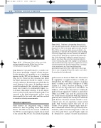
Figure 14.11 Problems in interpreting flow velocities
in a vein graft anastomosed to the posterior tibial artery. At point A, the PSV in the distal graft is 65 cm/s. At point B the PSV in the posterior tibial artery, just distal to the anastomosis, is 120 cm/s. This represents a near doubling in velocity, suggesting a stenosis. However, the diameter of the posterior tibial artery is significantly smaller than that of the distal graft, leading to a natural increase in systolic velocity, as flow velocity is inversely related to cross-sectional area, and in this example no narrowing is indicated. Unfortunately, at point C, a significant stenosis is demonstrated in the posterior tibial artery, 2 cm distal to the anastomosis, by color flow aliasing and a significant increase in the PSV, 400 cm/s. In this image there is some retrograde filling of the native vessel above the anastomosis (curved arrow).
C
A: Hyperemic flow is often seen in the early postoperative period. B: Over time, the flow
Figure 14.10
graft stenoses are shown in Table 14.2. Intervention by angioplasty or surgical revision is usually per- formed when the PSV ratio is equal to or greater than 3 (London et al 1993, Olojugba et al 1998, Landry et al 2002). Stenoses producing a PSV ratio of 2–2.9 are kept under close surveillance. In addition, a PSV of less than 45cm/s within the graft has been suggested to indicate a graft defect (Mills et al 1990). Care must be exercised in the use of this criterion as patients with large-diameter grafts may have relatively low velocity flow within the graft, because velocity is inversely related to cross- sectional area. In our experience, many normal grafts with velocities below this level do not occlude. Poor spectral Doppler angles may pro- duce considerable errors in one-spot velocity meas- urements, and it is therefore important to select an area of the graft where a good Doppler angle can be obtained for accurate velocity measurement.
