
Page 227 - Libro vascular I
P. 227
Chap-14.qxd 29~8~04 14:55 Page 218
218
PERIPHERAL VASCULAR ULTRASOUND
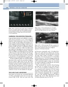
Figure 14.14 Extremely low volume flow recorded from an in situ vein graft indicates imminent graft occlusion. In this example the distal end of the graft had already thrombosed and the Doppler waveform demonstrates
no net forward flow.
COMMONLY ENCOUNTERED PROBLEMS
Large and obese patients can be difficult to examine, and it may be necessary to use a lower frequency transducer. Early postoperative scans can be difficult if the wounds are still healing, and scanning over a sterile transparent plastic dressing is useful in this sit- uation. Having no prior knowledge of the type and position of graft can lead to considerable problems. For example, a popliteal to tibial vein bypass graft may require the long saphenous vein to be harvested from the thigh, as it is larger at this level. Therefore, a large scar will be seen in the thigh, but the graft will not be located at this level; however, the sonogra- pher may automatically assume that this corresponds to the position of the proximal graft. A copy of the operation notes is a useful aid to locating the graft.
It is also possible for grafts to be routed in unusual directions, such as across the anterolateral thigh to join the anterior tibial artery in the calf. Some patients may have had a previous graft that has since occluded, and this could be mistaken for the new graft, which may still be patent. It is also possible for segments of native vessels to be patent, such as the superficial femoral artery, and this may cause some confusion or may even be mistaken for the graft.
TRUE AND FALSE ANEURYSMS
Vein grafts can develop true aneurysmal dilations over time, particularly at valve sites or at the anas- tomoses (Fig. 14.15). This can occur if the vein
Figure 14.15 An aneurysmal area in a vein graft corresponding to a valve site. Note the area of hyper- plasia or thrombus (arrow) in the area of dilation.
FA G
failure of the anastomosis. Note the corrugated appearance of the dacron material.
A false aneurysm (FA) has occurred at the distal end of a femorofemoral cross-over graft (G) due to
Figure 14.16
wall becomes structurally weak. A localized dou- bling in the graft diameter indicates the develop- ment of an aneurysm, and this should be reported and kept under regular surveillance to monitor progression. It is not uncommon to see thrombus in aneurysmal areas. Color flow imaging and spectral Doppler usually demonstrate areas of flow reversal in the aneurysmal regions. Large true aneurysms are repaired surgically by replacing the aneurysmal area with a new segment of vein.
False aneurysms are caused by blood flowing into and out of a defect in the vessel wall (see Ch. 11). They are typified as swirling areas of flow in a contained cavity outside the true flow lumen and may contain thrombus. They can occur if the suture line at the anastomosis fails or as a compli- cation of balloon angioplasty, due to splitting of the graft wall following high-pressure balloon inflation (Fig. 14.16). False aneurysms also occur at catheter puncture sites (see Ch. 11).
