
Page 30 - Libro vascular I
P. 30
ULTRASOUND AND IMAGING 21
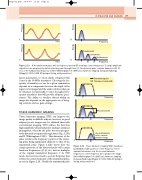
AB
Time f
CD
Time f
Frequency
Fundamental
1st harmonic
2nd harmonic
2f 3f Frequency
Figure 2.23 A: An undistorted pulse with its frequency spectra (B) showing a center frequency f. C: Large-amplitude signals become progressively distorted as they pass through tissue. D: The distorted pulse contains harmonics (2f, 3f, etc.) of the fundamental frequency f. (After Whittingham T A 1999 Tissue harmonic imaging. European Radiology 9(Suppl 3): S323–S326. © Springer-Verlag, with permission.)
better penetration, to 12 cm depth compared with 3 cm for the 10 MHz transducer. Choosing the fre- quency of transducer to use for a given examination depends on a compromise between the depth of the region to be imaged and the axial resolution that can be obtained. It is preferable to select the highest fre- quency transducer that will provide adequate pene- tration. The ability to visualize objects within an image also depends on the appropriate use of imag- ing controls, such as gain settings.
TISSUE HARMONIC IMAGING
A
B
C
f
f 2f
2f
Transmitted pulse Transducer band width
Frequency
Received echo
Frequency
Second harmonic echo—used to produce image
Frequency
Tissue harmonic imaging (THI) can improve the image quality in difficult subjects; however, in good subjects poorer images may be obtained than with conventional imaging. THI utilizes the fact that high-amplitude ultrasound pulses undergo nonlinear propagation, whereby the pulse becomes progres- sively distorted as it passes through tissue (Fig. 2.23A and B (Whittingham 1999)). This distortion of the pulse results in the frequency content of the return- ing pulse being significantly different to that of the transmitted pulse. Figure 2.23D shows how the energy spectrum of the distorted pulse will contain harmonic frequencies (2f, 3f, etc.) that are multiples of the original transmitted frequency, f. In THI mode, the receiver is tuned to a center frequency that is twice the center frequency of the transmitted pulse, as seen in Figure 2.24. Usually the transmitted pulse
Tissue harmonic imaging. Wide transducer bandwidths enable pulses of center frequency f to be
transmitted and use only the received harmonic frequencies, center frequency 2f, to produce the image. (After Whittingham T A 1999 Tissue harmonic imaging. European Radiology 9(Suppl 3): S323–S326. © Springer- Verlag, with permission.)
Figure 2.24
Chap-02.qxd 29~8~04 13:20 Page 21
Pressure Pressure
Energy Energy
