
Page 28 - Libro vascular I
P. 28
ULTRASOUND AND IMAGING 19
Beam-former delays
Beam-former delays
For receive beams
Receive beams
Transmission beam
A
Echo amplitude
B
C
Echo amplitude
Depth
Two scan line
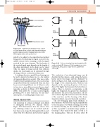
Figure 2.19 Parallel beam forming of two or more received beams from a single wide transmitted beam permits improvements in imaging frame rate. (After Whittingham 2003, with permission.)
path B to be added to the signal that has travelled along path A by delaying the signal received by the middle element before summing it with the signals received by the outer elements. The focal point of the received signal again depends on the lengths of the delays introduced. As the delay can be varied while the signal is being received from different depths, the focal length can be optimized through the image without a reduction in frame rate.
A technique known as parallel beam forming may be used to improve the frame rate (i.e., the number of images produced per second). This uses a wide, weakly focused transmitted beam. The received sig- nal produced from this transmitted beam can then be processed using different sets of delays in order to form two or more different received beams, simul- taneously, as shown in Figure 2.19 (Whittingham 2003). This allows two or more received signals, producing two or more scan lines, for each trans- mitted pulse, so enabling higher frame rates.
IMAGE RESOLUTION
The resolution of a system is defined as its ability to distinguish between two adjacent objects. Figure 2.20 demonstrates how the echoes from two reflecting surfaces can be resolved and also how they can no longer be distinguished from each other if the two objects are moved closer together.
Depth
Echoes returning from two boundaries (A) can be resolved (B). However, if the boundaries are close
together (C), they can no longer be seen as different echoes (D).
The resolution of an ultrasound image can be described in three planes—axial (along the beam), lateral (across the image) and slice thickness—as shown in Figure 2.21. Axial resolution depends on the length of the excitation pulse, which in turn depends on the operating frequency of the trans- ducer. The higher the frequency, the better the res- olution. There is, however, a compromise, as the higher the frequency, the greater the attenuation and therefore the poorer the penetration. The lat- eral resolution depends on factors such as the density of the scan lines and the focusing of the beam. Lateral resolution is poorer than axial resolution.
The out-of-imaging plane beam thickness, or slice thickness, will affect the region perpendicular to the scan plane over which returning echoes will be obtained. Ideally, the slice thickness should be as thin as possible to maintain image quality, so focusing is often used in this plane as well as in the imaging plane. This can be done either by
D
Figure 2.20
Chap-02.qxd 29~8~04 13:20 Page 19
