
Page 35 - Libro vascular I
P. 35
Chap-03.qxd 29~8~04 13:20 Page 26
26
PERIPHERAL VASCULAR ULTRASOUND
Back-scatter from blood
Blood is made up of red blood cells (erythrocytes), white blood cells (leukocytes) and platelets sus- pended in plasma. Red blood cells occupy between 36% and 54% of the total blood volume. They have a biconcave disc shape and a diameter of 7m, which is much smaller than the wavelength of ultrasound used to study blood flow. This means that groups of red blood cells act as scatterers of the ultrasound (see Fig. 2.8).
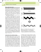
Transmitted signal voltage
A
Received signal voltage
B
Product
C
Doppler signal voltage
D
transmitted signal (A) by the received signal (B) and filtering out the high-frequency component (C) to leave the Doppler signal (D). (After Fish 1990, with permission.)
The back-scattered signal from blood received at the transducer is small, partly due to the back- scattered energy being radiated in all directions, unlike specular reflections, and partly because the effective cross-section of the blood cells is small compared with the width of the beam. The back- scattered power is proportional to the fourth power of the frequency (i.e., f 4), and therefore as the trans- mitted frequency selected to detect flow is increased, there is an increase in back-scattered power. How- ever, this is offset by the increase in attenuation of the overlying tissue with the increase in frequency. Ultrasound systems will often use a lower trans- mitted frequency for Doppler than for B-mode imaging, and the imaging and Doppler transmitted frequencies are usually indicated on the image. In situations in which blood velocity is low or blood cells are stationary, such as aneurysms or venous flow, the cells may aggregate into clumps, which can sometimes produce sufficiently high-amplitude back-scattered echoes to be displayed on the B-mode image (see Fig. 12.19).
EXTRACTING THE DOPPLER SIGNAL
The simplest Doppler systems consist of a trans- ducer with two piezoelectric elements (Fig. 3.2), one continuously transmitting ultrasound and the other continuously receiving back-scattered signals from both stationary tissue and flowing blood. This received signal therefore consists of both the trans- mitted frequency reflected by stationary objects and the Doppler-shifted frequencies back-scattered from moving blood cells. As the returning echoes are of low amplitude, first they must be amplified. The Doppler shift frequency can then be extracted from the received signal by a process known as demodulation. One method of demodulation used
multiplied by
equals
Time
Time
Time
Time
Filter
Demodulation. This is used to extract the Doppler frequency, in this case by multiplying the
Figure 3.4
in Doppler systems is shown in Figure 3.4. Here, the received signal is multiplied by the transmitted signal and the product is filtered to remove the high frequencies, thus providing the Doppler shift frequency. The received signal has a different fre- quency from the transmitted frequency, owing to the Doppler effect, and a lower amplitude, owing to attenuation of the signal by overlying tissue. As mentioned earlier, once the Doppler shift fre- quency has been extracted (by demodulation) and amplified, it can simply be output to a loudspeaker or investigated using a spectrum analyzer (Fig. 3.5). With experience, it is possible for the opera- tor to recognize the different sounds produced by normal and diseased vessels.
