
Page 55 - Libro vascular I
P. 55
Chap-04.qxd 29~8~04 13:22 Page 46
46
PERIPHERAL VASCULAR ULTRASOUND
the Doppler signal. If the color gain is increased to visualize the background noise, the operator will see the noise as a speckled pattern of all colors within the color box. This is because the noise generated within the scanner is a low-amplitude signal con- taining all frequencies. As the noise occurs in all frequencies, this noise is impossible to remove using the high-pass filter. As power Doppler dis- plays power rather than frequency, it is less suscep- tible to this low-amplitude noise since it is displayed as a darker color or not displayed at all.
The main disadvantage with power Doppler is that in order to improve sensitivity, a high degree of frame-averaging is used, which means that the operator has to keep the transducer still to obtain a good image. Therefore, this modality is less suit- able for rapidly scanning along vessels. The lack of angle dependence makes power Doppler useful in imaging tortuous vessels. Power Doppler also provides improved edge definition (e.g., around plaque). Some ultrasound systems provide a color flow display that combines the power Doppler dis- play with directional information. In this mode, the power of the signal is displayed as red for flow detected travelling toward the transducer, and the power of the signal detected from blood moving away from the transducer is displayed as blue. No velocity information is displayed in this mode.
ENHANCED FLOW IMAGING USING CONTRAST AGENTS AND HARMONIC IMAGING
A limiting factor in ultrasound imaging of flow is that the power of the ultrasound back-scattered from blood is much lower than that reflected from the surrounding tissue. Increasing the output power of the scanner will not overcome this problem as it would increase the signal from the surrounding tis- sue as well as from the blood. The concept behind the use of contrast agents in ultrasound is to intro- duce a substance into the blood that provides a higher back-scattered power than is available from blood alone. Contrast agents used clinically at present consist of microparticles to which gas microbubbles adhere. It is these microbubbles that provide the increase in back-scattered power. Contrast agents are divided into two types: right heart and left heart agents. Right heart agents are
A
Amplitude
B
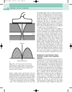
When the beam is at right angles to the blood flow, this will result in both negative and positive Doppler shift frequencies within the signal.
within a sample volume and therefore does not depend on whether the sample volume is totally or partially filled with the blood flow. Power Doppler is therefore able to provide better definition of the boundaries of the blood flow than color Doppler.
The improved sensitivity of the power Doppler is due to the relationship between the noise and
A: The beam used to detect the flow actually produces a range of angles of insonation. B:
Figure 4.16
Vessel
Doppler shift frequency
