
Page 53 - Libro vascular I
P. 53
Chap-04.qxd 29~8~04 13:22 Page 44
44
PERIPHERAL VASCULAR ULTRASOUND
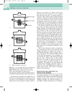
decrease in the frame rate. Figure 4.14C shows how the data from scan line 2 can be acquired while data from scan line 1 are being obtained, as scan line 1 will not detect pulses transmitted along scan line 2. The same is true for scan lines 3 and 4, and so forth. Extra lines of data can be created by averaging two adjacent lines to produce a scan line between them. As no new information is acquired to perform this, no change in the frame rate occurs.
The number of pulses used to produce each scan line of the color image is known as the ensemble length. Typically, an ensemble length of between 2 and 16 pulses is used to estimate the Doppler fre- quency. However, the more pulses that are used, the more accurate the estimate will be, and in situ- ations in which the returning Doppler signal is poor, a high number of pulses is required. There is, there- fore, a compromise between the accuracy of the fre- quency estimate and frame rate. The time taken for these 2 to 16 pulses to be transmitted and to return, the dwell time, obviously depends on the rate at which the pulses are transmitted (i.e., the PRF). When a low PRF is used, it will take longer for the pulse ensemble to be transmitted, leading to a lower frame rate.
These various limitations require a compromise to be made between the area over which the color Doppler information is acquired, the accuracy of the Doppler frequency estimate and the time it takes to acquire it. The selection of PRF, position of the ROI and frequency of the transducer are governed by the region of the body being imaged and the type of blood flow in that region. However, it is possible to optimize the frame rate by using as narrow an ROI as possible for the examination. The quality of the color image may be improved by averaging consecutive images, to reduce the noise, and displaying the image for a longer period of time. This control is sometimes known as the persistence.
RESOLUTION AND SENSITIVITY OF
COLOR FLOW IMAGING
The spatial resolution of the color image can be considered in three planes, as described for B-mode imaging (see Fig. 2.21). However, as blood flow imaging is dynamic, the temporal resolution (i.e., the ability to display changes that occur during a
Transducer
B-mode image
Color box ROI
Blood flow
A
B
12 34 56
C Order of acquisition of scan lines
Figure 4.14 The color image frame rate can be improved by (A) reducing the size of the color region of interest (ROI) or (B) reducing the density of the color scan lines.
(C) The scanner may improve the frame rate by interleaving the acquisition of data from different parts of the ROI. (After Ferrara & DeAngelis 1997, with permission.)
Interleaving the acquisition from different scan lines that are a distance apart can enable more than one pulse to be transmitted at a time, allowing an improvement in the frequency estimate without a
