
Page 52 - Libro vascular I
P. 52
Chap-04.qxd 29~8~04 13:22 Page 43
that produced by a higher PRF. It is therefore very important to select the appropriate PRF for the flow conditions to be imaged. If a low PRF is selected to image high-velocity flow, aliasing will occur, and if a high PRF is selected to image low-velocity flow, the flow may not be detected at all, as the dwell time will be too short (Fig. 4.9C). Ideally, a PRF should be selected that displays the highest velocities present with the colors near the top of the scale.
The cut-off frequency of the high-pass clutter filter will also affect the lowest frequencies that can be displayed. The high-pass filter will only allow frequencies greater than the cut-off frequency to be displayed, so that if this is set too high, the Doppler frequencies detected from the lower velocity blood flow will be removed. The level of the high-pass filter is usually displayed on the color scale (Fig. 4.13). Using the wrong filter setting has led to removal of the low velocities at the vessel walls or of low flow during diastole. The high-pass filter is linked to the PRF and therefore, as the PRF is increased, the high-pass filter is also automati- cally increased. However, some systems will allow the filter to be altered independently of the PRF, in which case the high-pass filter setting should be considered when the PRF is lower in order to image low-velocity flow.
FRAME RATE
The frame rate is the number of new images pro- duced per second. For color flow imaging to be
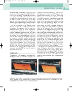
AB
Figure 4.13 Effect of using the filter. A: The filter is set too high, removing the low-velocity flow near the vessel walls (vertical arrows). B: The filter setting is reduced to display the low frequencies detected near the vessel walls. The filter setting may be displayed on the color scale (horizontal arrows).
CREATION OF A COLOR FLOW IMAGE
43
useful for visualizing pulsatile blood flow, a reason- ably high frame rate is required. With pulse echo imaging alone, the frame rate can be greater than 50 images per second. However, the time required to produce a color flow image is much longer and therefore the frame rates are much lower. The frame rate is dependent on several factors when using color flow imaging (Fig. 4.14). The ROI refers to the color box, which can be placed any- where within the image to examine blood flow. The size and position of the ROI have a significant effect on the frame rate. The width is especially important, as the wider the ROI, the more scan lines are required and therefore the longer it will take to collect the data for an image. The line den- sity (the number of scan lines per centimeter across the image) also affects the time taken to produce the image as the pulses for each scan line have to return before the next line can be produced. The length of the color box is less important. This is because the scanner has to wait for all the return- ing echoes before sending the next pulse, even if the information is not used to produce the image, so as not to suffer from range ambiguity.
The depth of the ROI is, however, an important factor. To image at depth, lower frequency ultra- sound is used, which will penetrate farther, allow- ing the ROI to be set at a greater depth. Therefore, the scanner will have to wait longer for the echoes to return from the greater depth and it will take longer to create each scan line, so reducing the frame rate.
