
Page 71 - Libro vascular I
P. 71
Chap-05.qxd 29~8~04 13:25 Page 62
62
PERIPHERAL VASCULAR ULTRASOUND
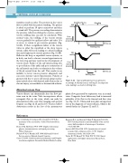
transducer and recorder. The pressure in the vein is first recorded with the patient standing. The patient is asked to perform 10 tiptoe maneuvers and then to stand still. The pressure recording demonstrates the pressure reduction during the exercise, and the venous refilling time can also be calculated. With normal veins, the refilling of the venous system occurs gradually by capillary inflow and takes 18s or more to return to pre-exercise pressures (Fig. 5.23A). If there is significant failure of the venous valves in either the superficial or the deep venous system, reflux will occur, leading to a shorter refilling time and a higher post-exercise pressure (Fig. 5.23B). Reflux in the deep or superficial venous systems, or in both, can lead to chronic venous hypertension in the lower leg and may result in the development of venous ulcers. Failure of the calf muscle pump due to poor flexion of the ankle and poor contraction of the calf muscle can lead to a reduction in the volume of blood ejected from the calf. This results in an inability to lower venous pressure adequately and can cause chronic venous hypertension. Patients at greatest risk due to poor calf muscle pump mecha- nism include those with limited ankle flexion due to chronic injury, osteoarthritis or rheumatoid arthritis.
Abnormal venous flow
Venous disease can dramatically alter the flow pat- terns seen in the veins. Valve incompetence allows retrograde flow in the veins, which can easily be demonstrated with color flow imaging and spectral Doppler (see Figs 12.15 and 12.16). Venous outflow obstruction results in the loss of the spontaneous
References
Caro C G, Pedley T J, Schroter R C, Seed W A 1978 The mechanics of the circulation. Oxford University Press, Oxford
Evans D H, McDicken W N 1999 Doppler ultrasound: physics, instrumentation, and signal processing. Wiley, Chichester
Nichols W N, O’Rourke M F 1990 McDonald’s blood flow in arteries. Edward Arnold, London
Oates C P 2001 Cardiovascular haemodynamics and Doppler waveforms explained. Greenwich Medical Media, London
Standing Tip-toe mmHg
Post- exercise pressure 0
A
100 mmHg
Post- exercise pressure
0
Standing
100
0
10 20
Refilling time
10 20
Refilling time
30 Time (s)
B
0
30 Time (s)
Typical ambulatory venous pressures recordings. A: Normal venous refilling. B: Incompetent
veins leading to a shorter refilling time.
Figure 5.23
phasic flow generated by respiration seen in normal veins. Congestive heart failure may lead to increased pulsatility of the flow in the femoral and iliac veins (see Fig. 13.17). Ultrasound now plays an important role in the diagnosis of venous disease, which is dis- cussed further in Chapters 12 and 13.
Reneman R S, van Merode T, Hick P, Hooks A P G 1985 Flow velocity patterns in and distensibility of the carotid artery bulb in subjects of various ages. Circulation 71(3):500–509
Spencer M P, Reid J M 1979 Quantitation of carotid stenosis with continuous-wave (C-W) Doppler ultrasound. Stroke 10(3):326–330
Taylor K J W, Burns P N, Wells P N T 1995 Clinical applications of Doppler ultrasound. Raven Press, New York
