
Page 73 - Libro vascular I
P. 73
Chap-06.qxd 29~8~04 14:41 Page 64
64
PERIPHERAL VASCULAR ULTRASOUND
ABC
Fast flow in center
Slow moving blood near vessel walls
DEF
Width of ultrasound beam
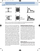
Figure 6.1 A, D: Velocity profiles for blunt flow and parabolic flow, respectively. B, E: If a wide ultrasound beam is used to insonate the vessel, all the velocities present will be detected. C, F: Idealised Doppler spectra that would be obtained from complete insonation of blunt flow and parabolic flow, respectively.
within the vessel will be detected and displayed on the spectrum. If blood is travelling with a blunt flow profile, most of the blood cells will be moving with the same velocity, and the spectrum will show only a small range of frequencies (Fig. 6.1A–C). If, however, the blood is travelling with a parabolic flow profile, then the blood in the center of the ves- sel will be travelling faster than that near the vessel walls and therefore the Doppler spectrum will dis- play a wide range of frequencies (Fig. 6.1D–F).
The spread of frequencies present within the spectrum at a given point in time is known as the degree of spectral broadening. Figure 6.1 shows the way in which the degree of spectral broadening depends on the velocity profile of the flow being interrogated, with greater spectral broadening seen in Figure 6.1F than in Figure 6.1C. The presence of turbulent flow (e.g., as a result of a stenosis) will increase spectral broadening, as the blood cells will be travelling with different velocities in random directions (see Fig. 5.20). Therefore, increased spec- tral broadening may indicate the presence of disease. However, the degree of spectral broadening can also be influenced by Doppler instrumentation, and this is known as intrinsic spectral broadening (discussed later in this chapter).
Nonuniform insonation of the vessel
The examples of idealized spectra given in Figure 6.1 assume that the beam evenly insonates the whole cross-section of the blood vessel in order to detect the correct proportions of all the blood velocities present. This is, however, an unrealistic situation as the Doppler beam can be quite narrow (of the order of 1 to 2 mm wide) and therefore may insonate only part of the artery or vein. If the beam passes through the center of the vessel (Fig. 6.2A), only part of the flow near the vessel walls (i.e., near the anterior and posterior walls) will be detected. The blood flow along the lateral walls will not be detected as it is not insonated by the Doppler beam. Therefore, in the presence of parabolic flow, the low-velocity flow near the walls will only be partially detected and the Doppler spectrum will no longer truly represent the low-velocity flow present within the vessel.
Sample volume size
The size and position of the sample volume, which can be controlled by the operator, will also affect the proportion of the vessel insonated. A small sample volume placed in the center of a large vessel
