
Page 75 - Libro vascular I
P. 75
Chap-06.qxd 29~8~04 14:41 Page 66
66
PERIPHERAL VASCULAR ULTRASOUND
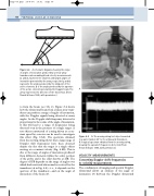
φ
θ1 θ2 θ3
Figure 6.4 A schematic diagram showing the range
of angles of insonation produced by a linear array transducer when making blood velocity measurements. 1 and 3 represent the smallest and largest angles of insonation generated by the array, respectively, and 2 represents the angle produced by the midpoint of the active elements. is the angle produced by the aperture of the active elements generating the Doppler beam. The arrow represents the direction of the blood flow. (From Thrush & Evans 1995, with permission.)
A
B
Figure 6.5 A: The moving string test object mounted in a water tank at 45° to the ultrasound transducer.
B: A typical spectrum obtained from the moving string, showing the spread of frequencies detected. (From Thrush & Evans 1995, with permission.)
to form the beam (see Ch. 2). Figure 6.4 shows how the ultrasound beam from a linear array trans- ducer can produce a range of angles of insonation, with the Doppler signals being detected at many angles. As the Doppler shift frequency detected is proportional to the cosine of the angle of insonation, , this will lead to a range of frequencies being detected even in the presence of a single target. A test object constructed of a string driven at a con- stant speed by a motor can be used to investigate this effect (Fig. 6.5A). The spectrum obtained from the moving string shows that a large range of Doppler shift frequencies have been detected despite the fact that the target is a single object moving at a constant velocity (Fig. 6.5B). This is due to the range of angles of insonation produced from different elements within the active portion of the probe and is the effect known as ISB. The degree of ISB depends on the range of angles over which back-scattered ultrasound is received by the transducer ( in Fig. 6.4)—i.e., it depends on the aperture of the transducer—and on the angle of insonation of the beam ().
VELOCITY MEASUREMENTS
Converting Doppler shift frequencies
to velocity measurements
The combination of imaging and spectral Doppler ultrasound allows an estimate of the angle of insonation () between the Doppler ultrasound
