
Page 74 - Libro vascular I
P. 74
Chap-06.qxd 29~8~04 14:41 Page 65
FACTORS THAT INFLUENCE THE DOPPLER SPECTRUM
65
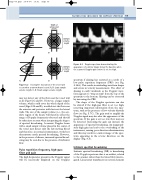
Width of ultrasound beam
AB
CD
Sample volume length
Sample volume length
Figure 6.2 Incomplete insonation of the vessel will occur when a narrow beam is used. A, B: Large sample volume length. C, D: Small sample volume length.
Figure 6.3 Doppler spectrum demonstrating the
appearance of a mirror image below the baseline when the scanner’s Doppler gain control is set too high.
spectrum if aliasing has occurred as a result of a low pulse repetition frequency (PRF) (see Fig. 3.14A). This results in misleading waveform shapes and errors in velocity measurement. The effect of aliasing is easily visualized, as the Doppler wave- form appears to ‘wrap around’ from the top of the spectrum to the bottom. Aliasing can be corrected by increasing the PRF.
The shape of the Doppler spectrum can also be altered if the high-pass filter is set too high, removing important information from the spec- trum, such as the presence of low-velocity diastolic flow (see Fig. 3.7C). The gain used to amplify the Doppler signal may also alter the appearance of the spectrum. If the gain is set too low, flow may not be detected. Increasing the gain can increase the appearance of spectral broadening. An inappropri- ately high gain can lead to the overloading of the instrument, causing poor direction discrimination, and this may result in a mirror image of the spec- trum appearing in the reverse direction on the display (Fig. 6.3).
Intrinsic spectral broadening
Intrinsic spectral broadening (ISB) is broadening of the Doppler spectrum that is an artifact, related to the scanner rather than the blood flow interro- gated. Linear array transducers use several elements
may not detect any of the flow near the vessel wall at all (Figs 6.2C and D). However, a larger sample volume, which could cover the whole depth of the vessel (Figs 6.2A and B), would detect the flow near the anterior and posterior walls but not the lateral walls. The size of the sample volume (i.e., the sen- sitive region of the beam) will therefore affect the range of Doppler frequencies detected and should be taken into account when interpreting the degree of spectral broadening. A narrow Doppler beam with a small sample volume placed in the center of the vessel may detect only the fast-moving blood and therefore, in normal circumstances, would not demonstrate much spectral broadening. However, in the presence of disease, increased spectral broad- ening may be seen due to the presence of turbulent flow.
Pulse repetition frequency, high-pass
filter and gain
The high frequencies present in the Doppler signal will be incorrectly displayed on the Doppler
