
Page 69 - Libro vascular I
P. 69
Chap-05.qxd 29~8~04 13:25 Page 60
60
PERIPHERAL VASCULAR ULTRASOUND
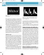
Figure 5.20 Doppler waveform demonstrating turbulent flow.
flow. In the presence of turbulence, not all the blood is travelling in the same direction, resulting in the angle of insonation being smaller for some parts of the blood flow. This results in turbulent spikes seen on the Doppler spectrum. It is possible for turbulent flow to occur only during the systolic phase of the cardiac cycle, when the systolic flow exceeds the crit- ical velocity and the diastolic flow does not.
The presence of turbulent flow causes energy to be lost, leading to an increased pressure drop across the stenosis. It is thought that bruits in the tissue near a stenosis (see Fig. 11.18A) may be due to perivascular tissue vibration caused by turbulence, and this may also lead to post-stenotic dilatation of the vessel. Vortices or irregular movement of a large portion of the fluid are more correctly referred to as disturbed flow rather than turbulent flow.
VENOUS FLOW
The venous system acts as a low-resistance pathway for blood to be returned to the heart. Veins are col- lapsible, thin-walled vessels capable of distending to a larger cross-sectional area than their corresponding arteries, so acting as a blood volume storage system which is important in the regulation of cardiac out- put. In addition, they also have a thermoregulation role in which blood is diverted to the superficial veins to reduce body temperature. The venous sys- tem can be divided into the central system (within the thorax and abdomen), the deep peripheral sys- tem and the superficial peripheral veins.
An important structural feature of the vein is the presence of very thin, but strong, bicuspid valves which prevent retrograde flow away from the heart. The vena cava and common iliac veins (see Fig. 12.3)
Figure 5.21 Doppler waveform showing the effect of changes in the pressure in the right atrium on blood flow in the jugular vein.
are valveless. Valves are found in the external iliac or common femoral veins in a proportion of the pop- ulation. Generally the more distal the vein, the greater the number of valves.
Venous flow back to the heart is influenced by respiration, the cardiac cycle and changes in posture.
Changes in flow due to the cardiac cycle
The central veins include the thoracic and abdominal veins, which drain to the right side of the heart via the inferior and superior venae cavae. The flow pat- tern and pressure in the central venous system are affected by changes in the volume of the right atrium, which occur during the cardiac cycle. Reverse flow occurs in the thoracic veins when the right atrium contracts, as there is no valve in the vena cava. This flow reversal can also be seen in the prox- imal veins of the arm and neck (Fig. 5.21) due to their proximity to the chest. During ventricular contraction, the atrium expands, increasing venous flow into the right atrium, and then flow gradually falls during diastole, only increasing briefly as the tri- cuspid valve opens. Flow patterns in the lower limb veins and peripheral arm veins are not significantly affected by the cardiac cycle due to vein compliance (which allows damping of the pressure changes), the presence of valves and changes in intra-abdominal pressure during respiration.
Effects of respiration on venous flow
Respiration has an important effect on venous pres- sure and flow because of changes in the volume of
