
Page 104 - Basic _ Clinical Pharmacology ( PDFDrive )
P. 104
90 SECTION II Autonomic Drugs
the ability to influence processes in distant regions of the body, and many clinical conditions. Unfortunately, a very large number of
extensive use of negative feedback. Both systems use chemicals for drugs used for other purposes (eg, allergies, mental illness) have
the transmission of information. In the nervous system, chemical unwanted effects on autonomic function.
transmission occurs between nerve cells and between nerve cells
and their effector cells. Chemical transmission takes place through
the release of small amounts of transmitter substances from the ANATOMY OF THE AUTONOMIC
nerve terminals into the synaptic cleft. The transmitter crosses the NERVOUS SYSTEM
cleft by diffusion and activates or inhibits the postsynaptic cell by
binding to a specialized receptor molecule. In a few cases, retrograde The ANS lends itself to division on anatomic grounds into two
transmission may occur from the postsynaptic cell to the presynap- major portions: the sympathetic (thoracolumbar) division
tic neuron terminal and modify its subsequent activity. and the parasympathetic (traditionally “craniosacral,” but see
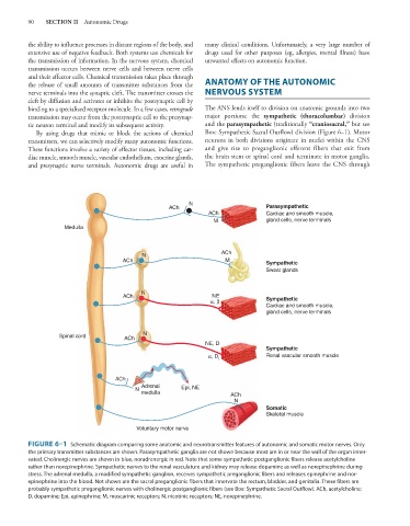
By using drugs that mimic or block the actions of chemical Box: Sympathetic Sacral Outflow) division (Figure 6–1). Motor
transmitters, we can selectively modify many autonomic functions. neurons in both divisions originate in nuclei within the CNS
These functions involve a variety of effector tissues, including car- and give rise to preganglionic efferent fibers that exit from
diac muscle, smooth muscle, vascular endothelium, exocrine glands, the brain stem or spinal cord and terminate in motor ganglia.
and presynaptic nerve terminals. Autonomic drugs are useful in The sympathetic preganglionic fibers leave the CNS through
N
ACh Parasympathetic
ACh Cardiac and smooth muscle,
M gland cells, nerve terminals
Medulla
N ACh
ACh M Sympathetic
Sweat glands
N
ACh NE Sympathetic
α, β
Cardiac and smooth muscle,
gland cells, nerve terminals
Spinal cord ACh N
NE, D
Sympathetic
α, D Renal vascular smooth muscle
1
ACh
Adrenal Epi, NE
N
medulla ACh
N
Somatic
Skeletal muscle
Voluntary motor nerve
FIGURE 6–1 Schematic diagram comparing some anatomic and neurotransmitter features of autonomic and somatic motor nerves. Only
the primary transmitter substances are shown. Parasympathetic ganglia are not shown because most are in or near the wall of the organ inner-
vated. Cholinergic nerves are shown in blue, noradrenergic in red. Note that some sympathetic postganglionic fibers release acetylcholine
rather than norepinephrine. Sympathetic nerves to the renal vasculature and kidney may release dopamine as well as norepinephrine during
stress. The adrenal medulla, a modified sympathetic ganglion, receives sympathetic preganglionic fibers and releases epinephrine and nor-
epinephrine into the blood. Not shown are the sacral preganglionic fibers that innervate the rectum, bladder, and genitalia. These fibers are
probably sympathetic preganglionic nerves with cholinergic postganglionic fibers (see Box: Sympathetic Sacral Outflow). ACh, acetylcholine;
D, dopamine; Epi, epinephrine; M, muscarinic receptors; N, nicotinic receptors; NE, norepinephrine.