
Page 105 - Basic _ Clinical Pharmacology ( PDFDrive )
P. 105
CHAPTER 6 Introduction to Autonomic Pharmacology 91
the thoracic, lumbar, and (according to new information) sacral nerves that are ontogenetically similar to sympathetic preganglionic
spinal nerves. The parasympathetic preganglionic fibers leave the fibers (see Box: Sympathetic Sacral Outflow). Note that the terms
CNS through the cranial nerves (especially the third, seventh, “sympathetic” and “parasympathetic” are anatomic designations
ninth, and tenth). and do not depend on the type of transmitter chemical released
Most thoracic and lumbar sympathetic preganglionic fibers are from the nerve endings nor on the kind of effect—excitatory or
short and terminate in ganglia located in the paravertebral chains inhibitory—evoked by nerve activity.
that lie on either side of the spinal column. Most of the remaining In addition to these clearly defined peripheral motor portions of
sympathetic preganglionic fibers are somewhat longer and termi- the ANS, large numbers of afferent fibers run from the periphery to
nate in prevertebral ganglia, which lie in front of the vertebrae, integrating centers, including the enteric plexuses in the gut, the auto-
usually on the ventral surface of the aorta. From the ganglia, post- nomic ganglia, and the CNS. Many of the sensory pathways that end
ganglionic sympathetic fibers run to the tissues innervated. Some in the CNS terminate in the hypothalamus and medulla and evoke
preganglionic parasympathetic fibers terminate in parasympathetic reflex motor activity that is carried to the effector cells by the efferent
ganglia located outside the organs innervated: the ciliary, pterygo- fibers described previously. There is increasing evidence that some of
palatine, submandibular, and otic ganglia. However, the majority these sensory fibers also have peripheral motor functions.
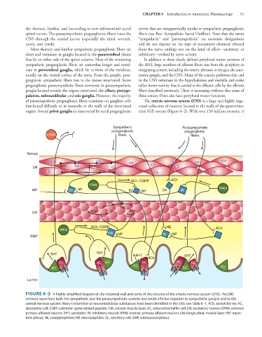
of parasympathetic preganglionic fibers terminate on ganglion cells The enteric nervous system (ENS) is a large and highly orga-
distributed diffusely or in networks in the walls of the innervated nized collection of neurons located in the walls of the gastrointes-
organs. Several pelvic ganglia are innervated by sacral preganglionic tinal (GI) system (Figure 6–2). With over 150 million neurons, it
Sympathetic Parasympathetic
postganglionic preganglionic
EPAN fibers fibers
Serosa
LM 5HT
ACh, CGRP ACh
IPAN
MP EN NP IN NP EN IN IPAN
NE
CM ACh ACh
ACh, CGRP NE ACh
IPAN
EN NP IN NP
SMP
5HT 5HT 5HT 5HT
ACh
SC AC EC SC AC SC
EC
EC EC EC
Lumen
FIGURE 6–2 A highly simplified diagram of the intestinal wall and some of the circuitry of the enteric nervous system (ENS). The ENS
receives input from both the sympathetic and the parasympathetic systems and sends afferent impulses to sympathetic ganglia and to the
central nervous system. Many transmitter or neuromodulator substances have been identified in the ENS; see Table 6–1. ACh, acetylcholine; AC,
absorptive cell; CGRP, calcitonin gene-related peptide; CM, circular muscle layer; EC, enterochromaffin cell; EN, excitatory neuron; EPAN, extrinsic
primary afferent neuron; 5HT, serotonin; IN, inhibitory neuron; IPAN, intrinsic primary afferent neuron; LM, longitudinal muscle layer; MP, myen-
teric plexus; NE, norepinephrine; NP, neuropeptides; SC, secretory cell; SMP, submucosal plexus.