
Page 33 - CJO_F17_GLAUCOMA_SUPPLEMENT
P. 33
MANAGING OPEN ANGLE GLAUCOMA
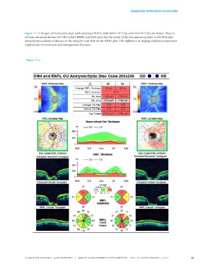
Figure 11: A 78 year-old Caucasian male with advanced POAG. Both RNFL OCT (a) and GCA OCT (b) are shown. There is
obvious advanced disease OS>OD on both RNFL and GCA plots but the extent of the loss appears greater in the GCA plot,
showing more advanced disease on the macular scan than on the RNFL plot. This difference in staging could have important
implications on treatment and management decisions.
Figure 11 a
CANADIAN JOURNAL of OPTOMETRY | REVUE CANADIENNE D’OPTOMÉTRIE VOL. 79 SUPPLEMENT 1, 2017 33