
Page 11 - herina surgery and possible lawsuits
P. 11
Anatomy
Inguinal region is the lower part of the anterolateral wall of abdomen and is limited
by the inguinal ligament inferiorly, lateral margin of rectus muscle medially and horizontal
line from anterior superior iliac spine to lateral border of rectus muscle superiorly. The word
“Groin” consists of the part 3cms above and 3cms below the inguinal ligament.
Anatomy of inguinal canal is of interest in understanding inguinal hernia.[36]
From superficial to deep the following layers are seen-fil]
1) Skin
2) Superficial fascia- it consists of two layers
a) Camper’s fascia- it is the fatty layer and it contains the cutaneous nerves
and vessels
b) Scarpa’s fascia- it is the membranous layer. Medially it is attached to the
linea alba and continue as the Codes’ fascia in the perineum. It passes over
the inguinal ligament and is continuous as the deep fascia of the thigh.
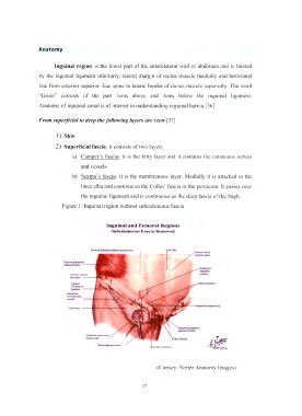
Figure 1: Inguinal region without subcutaneous fascia
In g u in a l a n d F e m o r a l R e g io n s
S u b c u ta n e o u s F asc ia R em o v ed
E t in n t l tb d o 4 rm » l o b hqu*
(•rrttfio f U9H)
E x ttm tl *6<J©fT*n*t
o t t q u * m u id #
A n ttf io t sup*fK>«
Am ---
In g u n *J
( P o u p w t'i)
hgtm+ni
c trc u m flM fliM
-Sup*fiic4«l mgurv^t nog
G«»«t * *ph+r>ou(
(Curtsey: Netter Anatomy Images)
17