
Page 15 - PR 2014 2016 02 Lasers Technology
P. 15
Lasers Technology | Progress Report 29
to kill bacteria, fungi, viruses, and protozoa, in- pH8) to remove non-adsorbed antigen. Then, a
cluding those resistant to conventional drugs. blocking solution is injected with the purpose
Alone, neither photosensitizer nor light pro- is of adhering in spaces of the channel where
duce damage in infected tissues. Studies are the antigen did not adhere. The entire loop is
performed in vitro and in vivo to investigate then washed again with TBS and inoculated
mechanisms and optimize PDI. Our results with primary antibody and subsequently with
show that PDI predominates on different tar- the secondary antibody, and finally washed
gets depending on cell growth phase. It can be again. A colorimetric reaction is produced in
enhanced by glucose and urea through differ- the microreactor to indicate the presence of
ent mechanisms , and induces programmed the antibody. The use of the development kit
cell death in protozoa, which contributes to whose substrate is orthophenylenediamine
reduce lesion size, parasite load and pain in showed a color change from transparent to
Leishmania amazonensis-induced cutaneous yellowish, evidencing the success of the device
leishmaniasis in mice. Besides, we designed a produced. Assays on plaques without the an-
dedicated light source to decontaminate bio- tigen were also made for the negative control.
medical instruments. In Veterinary Medicine,
PDI proved to be an alternative treatment for
caseous lymphadenitis abscesses in sheeps
and footpad dermatitis in penguins (Figure 8).
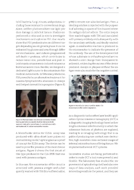
Figure 9: Microfluidic circuit used for ELISA. The
microreactor is the central serpentine.
As a diagnostic tool another laser health appli-
Figure 8: Photodynamic inactivation accelerates wound cation Optical coherence tomography (OCT) is
healing and reduces parasite load in cutaneous leish- a diagnostic imaging technology based on low
maniasis induced in paw of mice. A: control lesion with-
out treatment; B: lesion treated after 4 weeks. length coherence interferometry in which the
coherence features of photons are explored,
A Microfluidic device for ELISA assay was leading to an imaging technology that is ca-
produced with ultra-short laser pulses mi- pable of producing non-contact, non-destruc-
cromachining on BK7 optical glass as a proof tive, high-resolution cross-sectional images of
of concept for ELISA assay. The device can be internal microstructures of living tissues. We
used to prove the presence of the most diverse implemented several OCT systems.
antigens. Figure 9 shows the first circuit of
this type produced in the CLA-IPEN that was Innovative studies are being performed in
used with jararaca antigen. order to make OCT a tool more powerful and
flexible. The laboratory has studied the im-
In this case, the microreactor of the circuit is provement of optical setup itself and also new
sensitized with jararaca antigen and subse- ways of data analysis, such work provided
quently washed with TBS (tris-buffered saline, interesting results in the period, and they are