
Page 11 - PR 2014 2016 02 Lasers Technology
P. 11
Lasers Technology | Progress Report 25
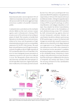
Diagnosis of skin cancer by ALA than MAL and according to LIF mea-
surements the PDT irradiation was performed,
Nonmelanoma skin cancers represent 95% of respectively, at 300 and 330 minutes after ALA
cutaneous neoplasms. Among them, squamous and MAL incubation. Histopathological analysis
cell carcinoma (SCC) is the more aggressive form evidenced necrosis and epithelial atrophy after
and shows a pattern of possible metastatic 10 days of PDT using both prodrugs, as well as
profile. reepitelization and collagen deposition at 20
days. Thus, despite the distinct concentration
5-aminolevulinic acid (ALA) and its methylat- of ALA and MAL used in the formulation of
ed ester (MAL) are the most common topical each photosensitizing cream, PDT mediated
agents used in photodynamic therapy (PDT) by both photosensitizing agents obtained
as precursors of the photosensitizer protopor- similar therapeutic outcomes. Besides, we
phyrin IX (PpIX). The induction of newly PpIX used Fourier transform infrared spectroscopy
depends on incubation time of each photosen- (FTIR) spectroscopy to assess the biochemical
sitizer in the tissue and the presence of high changes in normal skin caused by squamous
intralesional porphyrin levels is an important cell carcinoma induced by multi-stage chemical
parameter for the PDT effectiveness. We used carcinogenesis in mice. Changes in the absorp-
laser-induced fluorescence (LIF) spectroscopy to tion intensities and shifts were observed in
evaluate the optimum time to light exposure of the vibrational modes associated to proteins,
PDT mediated by ALA (20% w/w) and MAL (10% indicating changes in secondary conformation
w/w) to treat malignant lesions precursors of in the neoplastic tissue. Hierarchical cluster
cutaneous squamous cell carcinoma induced analysis was performed to evaluate the poten-
in mice. The therapeutic effects obtained by tial of the technique to differentiate the spectra
optimized ALA- and MAL-PDT were assessed 10 of neoplastic and normal skin tissue, so that
and 20 days after treatments. Higher PpIX levels the accuracy obtained for this classification
were evidenced in the lesions photosensitized was 86.4% (Figure 6).
A B
Figure 6: (a) Illustration of fluorescence from skin tumor treated by PDT mediated by ALA and MAL and histo-
logical results. (b) Discrimination of healthy and skin tumor by multivariate statistical analysis.