
Page 3 - Mesenchymal Stem Cell-Derived Exosomes as an Emerging Paradigm for Regenerative Therapy and Nano-Medicine
P. 3
Life 2021, 11, 784 3 of 26
release [20]. Although most studies on the molecular mechanism of exosome release are on
Life 2021, 11, x FOR PEER REVIEW 3 of 28
cancer, few (almost none) have reported on mesenchymal stem cell exosomes [21,22].
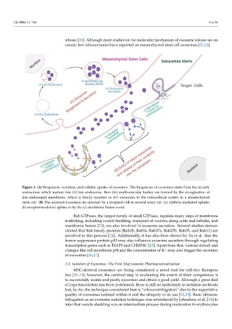
Figure 1. (A) Biogenesis, secretion, and cellular uptake of exosomes. The biogenesis of exosomes starts from the (i) early
Figure 1. (A) Biogenesis, secretion, and cellular uptake of exosomes. The biogenesis of exosomes starts from the (i) early
endosomes which mature into (ii) late endosome, then (iii) multivesicular bodies are formed by the invagination of late
endosomes which mature into (ii) late endosome, then (iii) multivesicular bodies are formed by the invagination of
endosomal membrane, which is finally secreted as (iv) exosomes to the extracellular matrix in a mesenchymal stem cell.
(B) The secreted exosomes are uptaken by a recipient cell in several ways viz. (a) clathrin‐mediated uptake, (b)
late endosomal membrane, which is finally secreted as (iv) exosomes to the extracellular matrix in a mesenchymal
receptor‐mediated uptake or by the (c) membrane fusion event.
stem cell. (B) The secreted exosomes are uptaken by a recipient cell in several ways viz. (a) clathrin-mediated uptake,
(b) receptor-mediated uptake or by the (c) membrane fusion event.
2.2. Exosome Secretion and Internalization
The release of exosomes into the extracellular milieu is governed by an orchestration
Rab GTPases, the largest family of small GTPases, regulate many steps of membrane
of proteins viz. soluble N‐ethylmaleimide‐ sensitive factor attachment protein receptors
trafficking, including vesicle budding, transport of vesicles along actin and tubulin, and
(SNAREs), tethering factors, Rabs, and other Ras GTPases [15]. The SNARE proteins, R‐
membrane fusion [23], are also involved in exosome secretion. Several studies demon-
or Q‐SNAREs, have been reported to affect exosome release. Fader et al. showed that the
strated that Rab family proteins (Rab2b, Rab5a, Rab27a, Rab27b, Rab35, and Rab11) are
R‐SNARE vesicle‐associated membrane protein 7 (VAMP7) is necessary for exosome
involved in this process [24]. Additionally, it has also been shown by Yu et al. that the
release in the human leukemic cell line K562 [16]. Another R‐SNARE protein, YKT6, is
tumor suppressor protein p53 may also influence exosome secretion through regulating
required for exosome release, as shown by two independent studies. Gross et al. showed
transcription genes such as TSAP6 and CHMP4C [25]. Apart from that, various stimuli and
that depletion of YKT6 decreased the level of TSG101, WNT3A, and VPS26/35 in
exosomes secreted from human embryonic kidney HEK293 cells [17]. Further,
changes like cell membrane pH and the concentration of K+ may also trigger the secretion
Ruiz‐Martinez et al. showed a reduced level of exosome‐associated TSG101 after the
of exosomes [26,27].
knockdown of YKT6 in A549 human lung cancer cells [18]. Similarly, in Drosophila S2
cells, depletion of the Q‐SNARE syntaxin 1A (Syx1A) decreased the release of EV
2.3. Isolation of Exosomes: The First Step towards Pharmaceuticalization
enriched v exosomes [19]. Wei et al. reported that pyruvate kinase type M2 (PKM2)
MSC-derived exosomes are being considered a novel tool for cell-free therapeu-
phosphorylates SNAP‐23, thus enabling exosome release [20]. Although most studies
tics [28–31]; however, the cardinal step in evaluating the extent of their competence is
on the molecular mechanism of exosome release are on cancer, few (almost none) have
to successfully isolate and purify exosomes and obtain a good yield. Although a great deal
reported on mesenchymal stem cell exosomes [21,22].
of experimentation has been performed, there is still no uniformity in isolation methods;
Rab GTPases, the largest family of small GTPases, regulate many steps of membrane
but, by far, the technique considered best is “ultracentrifugation” due to the superlative
trafficking, including vesicle budding, transport of vesicles along actin and tubulin, and
quality of exosomes isolated within it and the ubiquity of its use [32,33]. Basic ultracen-
trifugation as an exosome isolation technique was introduced by Johnstone et al. [34] to
infer that vesicle shedding was an intermediate process during maturation to erythrocytes.