
Page 66 - Mesenchymal Stem Cell-Derived Exosomes as an Emerging Paradigm for Regenerative Therapy and Nano-Medicine
P. 66
Extracellular Vesicle Treatment for Glaucoma IOVS j February 2018 j Vol. 59 j No. 2 j 706
Intravitreal Delivery of sEV
Under isoflurane-induced anesthesia, sEV were injected into
the vitreous, just posterior to the limbus using glass
micropipette. A 5-lL volume of sterile phosphate-buffered
9
saline (sPBS) loaded with 3 3 10 sEV was injected slowly and
the needle was retracted after a 1-minute delay to minimize
backflow. The concentration was chosen based on our
previous study 22 that demonstrated efficacy.
Electroretinography Measurements of the Positive
Scotopic Threshold Response
ERG was recorded using the Espion Ganzfeld full-field system
(Diagnosys LLC, Lowell, MA, USA) on day 0 before induction of
ocular hypertension, and on day 56/21 (Groups 2 and 3,
respectively) before animals were killed. Rats were dark
adaptedfor 12 hours overnight andpreparedfor ERG
recording under dim red light (>630 nm). Anesthesia was
induced with intraperitoneal injection of ketamine/xylazine
and eyes dilated with tropicamide. Scotopic flash ERG was
recorded from 5.5 to þ10 log units with respect to standard
flash in half log-unit steps. ERG traces were analyzed using in
built Espion software and the amplitude (with respect to
baseline) was used as a measure of rat visual function. Traces at
a light intensity of 1 3 10 5 mcd/s were chosen for analysis as
they gave a clean, unambiguous pSTR 100 ms after stimulus.
An individual masked to the treatment groups performed all
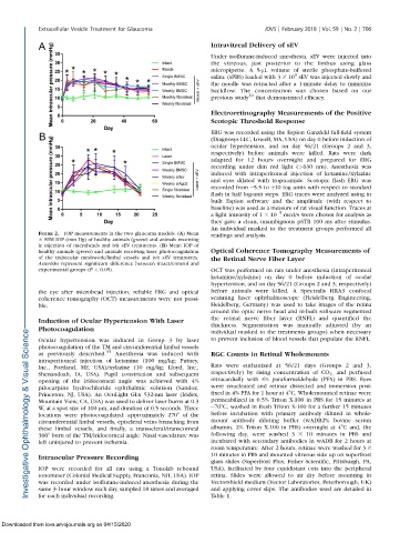
FIGURE 2. IOP measurements in the two glaucoma models. (A) Mean readings and analysis.
6 SEM IOP (mm Hg) of healthy animals (green) and animals receiving
ic injection of microbeads and ivit sEV treatments. (B) Mean IOP of
healthy animals (green) and animals receiving laser photocoagulation Optical Coherence Tomography Measurements of
of the trabecular meshwork/limbal vessels and ivit sEV treatments. the Retinal Nerve Fiber Layer
Asterisks represent significant difference between intact/control and
experimental groups (P < 0.05). OCT was performed on rats under anesthesia (intraperitoneal
ketamine/xylazine) on day 0 before induction of ocular
hypertension, and on day 56/21 (Groups 2 and 3, respectively)
the eye after microbead injection, reliable ERG and optical before animals were killed. A Spectralis HRA3 confocal
coherence tomography (OCT) measurements were not possi- scanning laser ophthalmoscope (Heidelberg Engineering,
ble. Heidelberg, Germany) was used to take images of the retina
around the optic nerve head and in-built software segmented
Induction of Ocular Hypertension With Laser the retinal nerve fiber layer (RNFL) and quantified the
thickness. Segmentation was manually adjusted (by an
Photocoagulation
individual masked to the treatments groups) when necessary
Ocular hypertension was induced in Group 3 by laser to prevent inclusion of blood vessels that populate the RNFL.
photocoagulation of the TM and circumferential limbal vessels
as previously described. 35 Anesthesia was induced with RGC Counts in Retinal Wholemounts
intraperitoneal injection of ketamine (100 mg/kg; Putney,
Inc., Portland, ME, USA)/xylazine (10 mg/kg; Lloyd, Inc., Rats were euthanized at 56/21 days (Groups 2 and 3,
Shenandoah, IA, USA). Pupil constriction and subsequent respectively) by rising concentration of CO 2, and perfused
opening of the iridocorneal angle was achieved with 4% intracardially with 4% paraformaldehyde (PFA) in PBS. Eyes
pilocarpine hydrochloride ophthalmic solution (Sandoz, were enucleated and retinae dissected and immersion post-
Princeton, NJ, USA). An OcuLight GLx 532-nm laser (Iridex, fixed in 4% PFA for 1 hour at 48C. Wholemounted retinae were
Mountain View, CA, USA) was used to deliver laser burns at 0.3 permeabilized in 0.5% Triton X-100 in PBS for 15 minutes at
W, at a spot size of 100 lm, and duration of 0.5 seconds. Three 708C, washed in fresh Triton X-100 for a further 15 minutes
locations were photocoagulated: approximately 2708 of the before incubation with primary antibody diluted in whole-
circumferential limbal vessels, episcleral veins branching from mount antibody diluting buffer (wADB2% bovine serum
these limbal vessels, and finally, a transscleral/transcorneal albumin, 2% Triton X-100 in PBS) overnight at 48C and, the
3608 burn of the TM/iridocorneal angle. Nasal vasculature was following day, were washed 3 3 10 minutes in PBS and
left uninjured to prevent ischemia. incubated with secondary antibodies in wADB for 2 hours at
room temperature. After 2 hours, retinae were washed for 3 3
10 minutes in PBS and mounted vitreous side up on superfrost
Intraocular Pressure Recording
glass slides (Superfrost Plus; Fisher Scientific, Pittsburgh, PA,
IOP were recorded for all rats using a Tonolab rebound USA), facilitated by four equidistant cuts into the peripheral
tonometer (Colonial Medical Supply, Franconia, NH, USA). IOP retina. Slides were allowed to air dry before mounting in
was recorded under isoflurane-induced anesthesia during the Vectorshield medium (Vector Laboratories, Peterborough, UK)
same 3- hour window each day, sampled 18 times and averaged and applying cover slips. The antibodies used are detailed in
for each individual recording. Table 1.
Downloaded from iovs.arvojournals.org on 04/15/2020