
Page 67 - Mesenchymal Stem Cell-Derived Exosomes as an Emerging Paradigm for Regenerative Therapy and Nano-Medicine
P. 67
Extracellular Vesicle Treatment for Glaucoma IOVS j February 2018 j Vol. 59 j No. 2 j 707
þ
þ
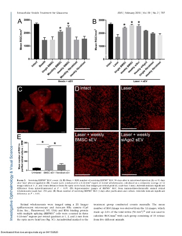
FIGURE 3. Surviving RBPMS RGC count. (A, B) Mean 6 SEM number of surviving RBPMS RGC 56 days after ic microbead injection (A)or 21 days
2
after laser photocoagulation (B). Counts were conducted in a 0.33-mm region of retinal wholemounts, calculated as a composite average of 12
images taken at 1-, 2-, and 3-mm distances from the optic nerve head, four images per retinal petal (C; scale bar, 1 mm). Asterisks indicate significant
difference from injured/untreated at P < 0.05. (D) Representative images of RBPMS þ RGC from immunohistochemically stained retinal
þ
wholemounts (scale bar: 250 lm). (E) Mean number of surviving RBPMS RGC 3 days after purification and culture. Asterisks indicate significant
difference at P < 0.01.
Retinal wholemounts were imaged using a Z1 Imager treatment group conducted counts manually. The mean
epifluorescent microscope and Axiocam HRc camera (Carl number of RGC/image was derived from the 12 images, which
Zeiss, Inc., Thornwood, NY, USA) and RNA binding protein made up 2.6% of the total retina (50 mm ) and was used to
2 36
þ
with multiple splicing (RBPMS) cells were counted in three 2
2
0.33-mm regions per retinal quadrant at 1, 2, and 3 mm from calculate RGC/mm with each group consisting of 10 retinae
the optic nerve head (see Fig. 3C). An individual masked to the from five different animals.
Downloaded from iovs.arvojournals.org on 04/15/2020