Page 28 - SRL Diagnostics
P. 28

TECHNOLOGIES & PLATFORMS-CYTOGENETICS
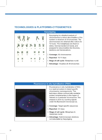
 Page 12                                        Karyotyping
                                                     Karyotyping is a detailed analysis of
                                                     chromosomes to detect abnormalities of the
                                                     number or structure of chromosomes. The
                                                     chromosomes are obtained after culture for
                                                     72 hours. The metaphases are fixed on
                                                     slides, Giemsa banded (G-band), and
                                                     analyzed for abnormalities like trisomies,
                                                     deletions and translocations.

                                                     Coverage: All chromosomes
                                                     Reported: 10-14 days
                                                     Stage of cell cycle: Metaphase nuclei       Page 22                                                     Page 26
                                                     Advantage: Visualize all chromosomes


                                     Fluorescence in situ hybridization (FISH)
                                                     Fluorescence in situ hybridization (FISH)
                                                     is a rapid procedure to detect specific
                                                     chromosome rearrangements. This
                                                     technique utilizes commercially available
                                                     probes complementary to the region of
                                                     interest on a particular chromosome. The
                                                     analysis is done by counting signals
                                                     under the fluorescent microscope as.

                                                     Coverage: Target specific sequences
                                                     Reported: 3-5 days
                                                     Stage of cell cycle: Metaphase &
                                                     Interphase nuclei
                                                     Advantage: Submicroscopic deletions
                                                     not detectable by Karyotyping


           25
   23   24   25   26   27   28   29   30   31   32   33