
Page 295 - Medicine and Surgery
P. 295
P1: FAW
BLUK007-07 BLUK007-Kendall May 25, 2005 18:18 Char Count= 0
Chapter 7: Clinical 291
Increased tone (spasticity). Decreased or absent reflexes. Plantars remain down-
Decreased power in a pyramidal distribution (i.e. af- going (or are absent).
fecting extensors more than flexors in the upper limbs, Wasting develops within 3 weeks of a lesion.
but affecting flexors more than extensors in the lower Fasciculations, which are small local contractions of
limbs). muscle motor units, due to spontaneous discharge of
Increased tendon reflexes, absent abdominal reflexes muscle fibres innervated by a single motor nerve fila-
and up-going (extensor) plantar reflexes. ment.
No muscle wasting or fasciculations (wasting may oc-
cur in long standing lesions due to disuse atrophy).
Patterns of LMN weakness
The pattern depends on which nerves or roots are af-
Patterns of UMN weakness
fected, and at what level.
Depending on the severity, the weakness may be de- Anterior horn cell lesions occur as part of motor neu-
scribed as a ‘plegia’ = total paralysis, or a ‘paresis’ =
rone disease, polio or other viral infections, and can
partial paralysis, but these terms are often used inter-
affect multiple levels.
changeably (see Table 7.3).
Spinal root damage is often due to compression for
Cerebral hemisphere disease may occur either in the
example, a disc protrusion causing compression at T1
cortex or the internal capsule. Common causes are st-
will cause weakness and wasting of the small muscles
rokes(vascularocclusionorhaemorrhage)andtumours.
of the hand.
Internal carotid artery occlusion may cause a hemi-
The brachial and lumbosacral plexus can be affected
paresis.
by inflammation or trauma.
Occlusion of the middle cerebral artery territory may
Singleormultipleperipheralnervelesionscauseweak-
cause UMN signs more in the arms than the legs.
ness in the distribution of that nerve or as part of a
Occlusion of the anterior cerebral artery (ACA) terri-
multiple neuropathy.
tory may cause UMN signs more in the legs than the
arms.
Patterns of sensory loss
Lower motor neurone signs
Cerebellar signs
Lower motor neurone (LMN) signs are due to lesions of
the anterior horn cell (or cranial nerve nucleus), the mo- Nystagmus: This is usually horizontal and the fast
tornerverootleavingthespinalcord,oroftheperipheral phase is towards the side of the lesion.
nerves): Dysarthria:Scanningspeech,whichiswhenthespeech
Decreased tone (flaccidity). is heard syllable by syllable. Ask the patient to say
Decreased power in the distribution of the affected ‘British Constitution’ or ‘West Register Street’. It oc-
nerves or nerve roots (not pyramidal). curs when both lateral lobes are affected.
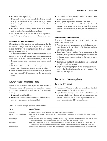
Table 7.3 Patterns of upper motor neurone weakness
Term Area of lesion
Diplegia A type of cerebral palsy affecting the lower limbs predominantly.
Quadriplegia/tetraplegia Affecting all four limbs. Usually due to a cervical spinal cord lesion, occasionally bilateral cerebral
lesions.
Hemiplegia Weakness of one half of the body (sometimes including the face) caused be a contralateral cerebral
hemisphere lesion, a brainstem lesion or ipsilateral spinal cord lesion (unusual).
Paraplegia Affecting both lower limbs, and usually caused by a thoracic or lumbar spinal cord lesion e.g. cord
compression. Bilateral hemisphere (anterior cerebral artery) lesions can cause this but are rare.
Monoplegia Contralateral hemisphere lesion in the motor cortex causing weakness of one limb, usually the arm.