
Page 66 - Science
P. 66
RESEARCH | RESEARCH ARTICLE
enrichment of the anchor pfam15611, represent- SECphi27, and lambda-vir phages (Figs. 2C and 3, and motB, respectively (Fig. 3C). MotA and MotB
ing a domain of unknown function, within de- A and C, and fig. S3). Further searches based on are inner membrane proteins that are part of
fense islands. Pfam15611-containing gene clusters homologies to the first two genes of the system, the flagellar motor of bacteria. They assemble
were previously reported as genomically asso- zorA and zorB, revealed a second type of Zorya, into a MotAB complex, which forms the stator
ciated with tellurium- and stress-resistance genes comprised of the three genes zorABE.Atype II of the flagellar motor (the static part within
(23). The reconstructed system is composed of the Zorya was cloned from E. coli ATCC8739 into which the flagellar rotor swivels) (24). The MotAB
four genes zorABCD, overall encompassing ~9 kb E. coli MG1655 and provided defense against T7 complex also forms the proton channel that
of DNA, with pfam15611 being the third gene in and the ssDNA phage SECphi17 (Figs. 2C and 3, B provides the energy for flagellar rotation, cou-
the system (zorC) (Fig. 3C and Table 1). A repre- and C, and fig. S3). pling transport of protons into the cell with the
sentative Zorya operon from E. coli E24377A was The first two genes of the Zorya system, zorA rotation (Fig. 3D) (25, 26). Whereas zorB shares
cloned into E. coli MG1655 and provided 10- to and zorB, contain protein domains sharing dis- the same size and domain organization with
10,000-fold protection against infection by T7, tant, but clear, homology with domains in motA motB (including the pfam13677 and pfam00691 Downloaded from
http://science.sciencemag.org/
on March 1, 2018
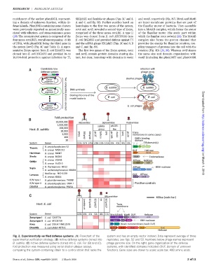
Fig. 2. Experimentally verified defense systems. (A) Flowchart of the system and has an empty vector instead. Data represent average of three
experimental verification strategy. (B) Active defense systems cloned into replicates; see figs. S2 and S3. Numbers below phage names represent
B. subtilis.(C) Active defense systems cloned into E. coli. For (B) and (C), phage genome size. On the right, gene organization of the defense
fold protection was measured using serial dilution plaque assays, systems, with identified domains indicated (DUF, domain of unknown
comparing the system-containing strain to a control strain that lacks the function). Gene sizes are drawn to scale; scale bar, 400 amino acids.
Doron et al., Science 359, eaar4120 (2018) 2 March 2018 3of 11