
Page 103 - Current techniques in canine and feline neurosurgery_2017_Neat
P. 103
A B
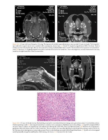
Figure 10.5 A 9‐year‐old female Hungarian Vizla dog. The dog was referred after a generalized seizure was recorded 24 hours previously. The histopathol
ogy results after surgical removal were consistent with a meningioma, atypical type. (A) Dorsal T1‐weighted postgadolinium MRI of the brain. There is
evidence of a mass affecting the right olfactory and frontal lobe causing a marked midline shift to the left. This mass shows heterogeneous contrast enhance
ment. (B) Transverse T1‐weighted postgadolinium MRI of the brain at the level of the frontal lobe. There is heterogeneous contrast enhancement of the mass
located on the right frontal lobe. There is a mass effect.
A B
C
Figure 10.6 A 12‐year‐old female German Shepherd dog presented for intermittent behavioral changes and a generalized seizure 2 months before referral.
The histopathology results (C) after surgical removal were consistent with a meningioma, fibroblastic microcystic subtype. (A) Sagittal T2‐weighted MRI of
the brain. There is evidence of a hyperintense well‐defined mass affecting the frontal lobe. There is perilesional edema. (B) Transverse T1‐weighted post
gadolinium MRI of the brain at the level of the frontal lobe. There is evidence of a mass effect affecting the right frontal lobe causing a midline shift to the
left. This mass shows heterogeneous contrast enhancement. There is a dural tail sign associated with the longitudinal fissure. (C) Spindle cells with elongated
nuclei haphazardly arranged in small bundles. H&E staining, ×20. Source: (C) Courtesy of Dr. Jim Cooley.