
Page 104 - Current techniques in canine and feline neurosurgery_2017_Neat
P. 104
102 Section II: Intracranial Procedures
Vascular (frontotemporal craniotomy) in combination with antifungal ther
Hemorrhage on the frontal lobe which necessitates surgical apy remains the treatment of choice to improve survival in CNS
decompression is uncommon. However, if the neurological symp aspergillosis [21].
toms are severe, persist, or deteriorate, transfrontal craniotomy to
allow decompression might be required [15]. The frontal lobe is Malformation
supplied by the rostral and middle cerebral arteries (see Figure 10.1). Congenital malformations affecting the frontal lobe cortex include:
Rupture of a blood vessel wall (artery or vein) causing hemorrhage anencephaly, meningocele, exencephaly, lipomeningocele, and holo
can be classified as primary or secondary depending on the under prosencephaly–arrhinencephaly. Meningoencephalocele is a protru
lying cause of bleeding. Primary hemorrhage originates from the sion of cerebral tissue and meninges through a congenital defect in
spontaneous rupture of small damaged vessels, while secondary the cranial bones (cranioschisis or cranium bifidum) whereas hernia
hemorrhage has been reported in dogs in association with various tion of only meninges is a meningocele [22]. Meningoencephalocele
causes, such as rupture of congenital vascular abnormalities, hem is the most common of these two malformations, but microscopic
orrhage into brain tumors, inflammatory disease, brain infarction, examination is commonly required to appreciate the difference.
or impaired coagulation [16]. Extradural, subdural, epidural, suba Generalized seizures not responding to medical treatment is the
rachnoid, and intraparenchymal hemorrhage has been reported in main neurological sign when the meningocele only affects the fron
dogs and cats following head injury [15,17,18]. tal lobe [23,24]. Transfrontal craniotomy with excision of a menin
goencephalocele and closure of the dural defect was an effective
Trauma treatment for an intranasal meningoencephalocele in a dog, and was
The frontal lobe is protected by the skull and the frontal sinus (see described by Martlé et al. [24].
Figures 10.2 and 10.3). However, penetrating trauma can reach the
frontal lobe [15,19] through the frontal sinus and cribriform plate
(Figure 10.7). If the neurological signs are severe and deteriorate sec Surgical Technique (Video 10.1)
ondary to a skull depression, hemorrhage, or a hypertensive pneumo Several techniques have been described [10,11,13,14]. The tech
cephalus, transfrontal craniotomy might be indicated [15,20]. niques vary by the region (olfactory vs. frontal lobes) and the type
of intervention necessary. The approach to the frontal lobe and
olfactory bulb is generally approached via the diamond‐shaped or
Infectious
Frontal lobe abscess or empyema, are often secondary to head trauma trapezoidal‐shaped bone flap over the rostral extent of the frontal
or migrating foreign bodies (Figure 10.8). A transfrontal approach bone sinus [13,14]. Bilateral and unilateral approaches have been
might be needed to decompress, debride and culture suppurative described. However, the unilateral approach has limited visibility
material [20]. Access to a more ventral empyema might not be and restricted access to the frontal and olfactory lobes.
achieved with this technique (Figure 10.8). Nevertheless, sample col The dog is positioned in sternal recumbency; the head can be
lection for culture or biopsy (ultrasound guided if needed) is possible raised by air cushions or sand bags and the table inclined so the head
through the transfrontal approach.
Frontal lobe extension from aspergillosis is uncommon in veteri
nary medicine. However, in humans, neurosurgical intervention
Figure 10.8 A 5‐year‐old female Domestic Shorthair cat that was referred
for depression, coughing, and retching of several weeks’ duration. The refer
ring veterinarian had extracted two fragments of grass awn from the oro
pharynx. Dorsal T1‐weighted postgadolinium MRI of the brain shows a
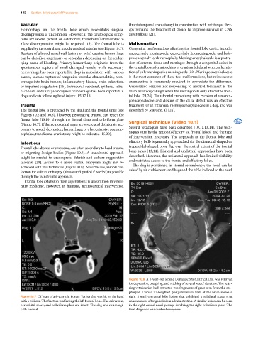
Figure 10.7 CT scan of a 9‐year‐old Border Terrier that was hit on the head right frontal–temporal lobe lesion that exhibited a subdural space ring
with a pickaxe. The fracture is affecting the left frontal bone. The calvarium, enhancement after gadolinium administration. A similar lesion can be seen
periorbital space, and cribriform plate are intact. The dog was neurologi in the right caudal nasal passage involving the right cribriform plate. The
cally normal. final diagnosis was cerebral empyema.