
Page 51 - Current techniques in canine and feline neurosurgery_2017_Neat
P. 51
Chapter 4: Advanced Imaging: Intracranial Surgery 43
may offer some diagnostic benefit for ischemic stroke, open in toy breed dogs with congenital hydrocephalus and this
especially in cases that could be confused with inflammatory allows ultrasonography of the brain to identify the enlarged
lesions [115]. lateral anechoic ventricles [5,6]. However, this is not always the
Radiography and ultrasonography are useful in looking for the case especially if the hydrocephalus is not a congenital lesion.
underlying systemic causes of strokes (renal disease and adrenal Although the diagnosis may be made with ultrasound in some
disease most commonly) [112]. cases, MRI is indicated to confirm the diagnosis and identify
any underlying cause for the hydrocephalus. In most cases of
Head Trauma congenital hydrocephalus, only the lateral ventricles, with or
Even mild neurological signs following head trauma exhibit a without the third ventricle, is affected. Dilation of the mesen-
high incidence of lesions detectable on CT [116]. Most cases of cephalic aqueduct and fourth ventricle often indicates obstruc-
head trauma resulting in neurological signs will show changes on tion to CSF flow at the lateral apertures of the fourth ventricle
MRI and the imaging study needs to determine if there is signifi- or the foramen magnum. Mild hydrocephalus is commonly
cant brain compression due to fracture fragments or hematoma seen in association with occipital malformation (Chiari‐like
formation or mass effect. CT examination will clearly show the malformation) but intracranial signs are not usually seen. In
presence of skull fractures and allow detection of depressed frac- older animals, hydrocephalus is often secondary to inflamma-
tures that may need surgical decompression. MRI, while not tory or neoplastic disease. In such cases, it is essential that
showing such clear bone detail, provides more information on FLAIR images be obtained to identify periventricular lesions.
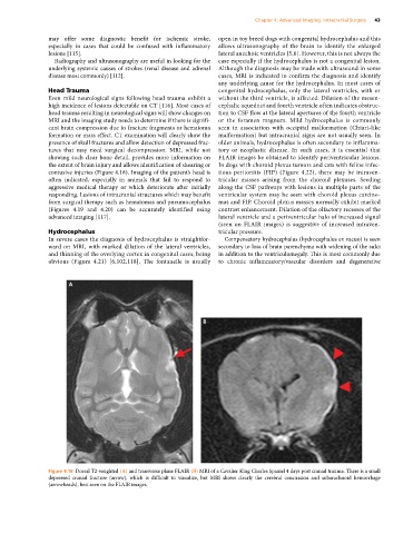
the extent of brain injury and allows identification of shearing or In dogs with choroid plexus tumors and cats with feline infec-
contusive injuries (Figure 4.18). Imaging of the patient’s head is tious peritonitis (FIP) (Figure 4.22), there may be intraven-
often indicated, especially in animals that fail to respond to tricular masses arising from the choroid plexuses. Seeding
aggressive medical therapy or which deteriorate after initially along the CSF pathways with lesions in multiple parts of the
responding. Lesions of intracranial structures which may benefit ventricular system may be seen with choroid plexus carcino-
from surgical therapy such as hematomas and pneumocephalus mas and FIP. Choroid plexus masses normally exhibit marked
(Figures 4.19 and 4.20) can be accurately identified using contrast enhancement. Dilation of the olfactory recesses of the
advanced imaging [117]. lateral ventricle and a periventricular halo of increased signal
(seen on FLAIR images) is suggestive of increased intraven-
Hydrocephalus tricular pressure.
In severe cases the diagnosis of hydrocephalus is straightfor- Compensatory hydrocephalus (hydrocephalus ex vacuo) is seen
ward on MRI, with marked dilation of the lateral ventricles, secondary to loss of brain parenchyma with widening of the sulci
and thinning of the overlying cortex in congenital cases, being in addition to the ventriculomegaly. This is most commonly due
obvious (Figure 4.21) [6,102,118]. The fontanelle is usually to chronic inflammatory/vascular disorders and degenerative
A
B
Figure 4.18 Dorsal T2‐weighted (A) and transverse plane FLAIR (B) MRI of a Cavalier King Charles Spaniel 4 days post cranial trauma. There is a small
depressed cranial fracture (arrow), which is difficult to visualize, but MRI shows clearly the cerebral concussion and subarachnoid hemorrhage
(arrowheads), best seen on the FLAIR images.