
Page 55 - Current techniques in canine and feline neurosurgery_2017_Neat
P. 55
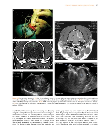
Chapter 4: Advanced Imaging: Intracranial Surgery  47


                A


                                                                 D


                B


                                                                 E


                C


               Figure 4.24  Intraoperative ultrasound. (A) The ultrasound probe (arrow) is covered with a sterile sleeve, the opening in the calvarium is flooded with
               warm saline, and the probe is placed directly on the brain parenchyma. Sterile ultrasound gel is placed inside the sleeve. (B, C) A transfalcine meningioma
               on CT and intraoperatively using ultrasound. (D, E) A left cortical high‐grade glioma on transverse MRI and on intraoperative ultrasound imaging.
               In (E), the asterisk denotes the hyperechoic mass and the star a hypoechoic region where some of the mass has been removed using an ultrasonic surgical
               aspirator (CUSA).


               was  identified  intraoperatively  after  craniectomy  and  durotomy   cerebri, gray  matter,  and  white  matter  were  easily  differentiated
               using a 5–8 MHz probe enveloped in a sterile sleeve containing ster-  from the generally more heterogeneous hyperechoic intracranial
               ile ultrasound lubricant. The covered probe was placed directly on   tumors. Cystic areas of the tumors and the one cystic mass in this
               the cerebral, cerebellar, or brainstem tissue to visualize the mass   study were contrasted from surrounding structures by their
               after flooding the craniectomy site with sterile saline. The intracra-  hypoechogenicity. Size and shape of the masses approximated the
               nial lesions were imaged in both sagittal and transverse planes.   same parameters  identified on advanced imaging (MRI,  CT).
               Most of  each intracranial  neoplasm  appeared distinctly hypere-  Noncystic portions of the meningiomas appeared most hypere-
               choic. The distinct imaging characteristics of the ventricles, falx   choic. The gliomas often were less heterogeneously hyperechoic