
Page 73 - Current techniques in canine and feline neurosurgery_2017_Neat
P. 73
A B
C
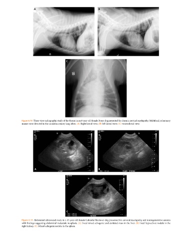
Figure 6.10 Three‐view radiographic study of the thorax in an 8‐year‐old female Boxer dog presented for chronic cervical myelopathy. Multifocal pulmonary
masses were detected in the caudal/accessory lung lobes. (A) Right lateral view; (B) left lateral view; (C) ventrodorsal view.
A B
C
Figure 6.11 Abdominal ultrasound study in a 13‐year‐old female Labrador Retriever dog presented for cervical myelopathy and nonregenerative anemia
with findings suggesting abdominal metastatic neoplasia. (A) Focal mixed echogenic and cavitated mass in the liver. (B) Focal hypoechoic nodule in the
right kidney. (C) Mixed echogenic nodule in the spleen.