
Page 74 - Current techniques in canine and feline neurosurgery_2017_Neat
P. 74
68 Section I: Diagnostics and Planning
A
B
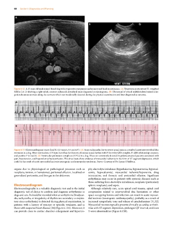
Figure 6.12 A 13‐year‐old male mixed‐breed dog with progressive paraparesis and urinary and fecal incontinence. (A) Transverse postcontrast T1‐weighted
MRI at L4–L5 showing a right‐sided, contrast‐enhanced, intradural mass diagnosed as meningioma. (B) Ultrasound of a focal multilobulated mixed echo-
genicsubcutaneous mass along the sternum which was incidentally detected during the physical examination and later diagnosed as sarcoma.
A
B
Figure 6.13 Electrocardiogram traces (lead II, 12.5 mm/s, 0.5 cm/mV). (A) Sinus tachycardia due to severe spinal pain as a result of acute intervertebral disc
extrusion in a dog. Heart rate is about 170 bpm but it has the features of normal sinus rhythm with P for every QRS complex, P–QRS relationship constant,
and positive P in lead II. (B) Ventricular premature complexes (VPCs) in a dog. These are commonly detected in polytraumatized patients associated with
pain, hypotension, and hypovolemia/hypoperfusion. The sinus beats show evidence of myocardial ischemia in the form of ST‐segment depression, which
could be the result of acute myocardial necrosis/neurogenic cardiomyopathy syndrome. Source: Courtesy of Dr. Lynne O’Sullivan.
organs due to physiological or pathological processes such as phy, electrolyte imbalance (hyperkalemia, hyponatremia, hypercal-
neoplasia, torsion, or hematoma), peritoneal effusion, localized or cemia, hypocalcemia), myocardial ischemia/hypoxemia, drug
generalized peritonitis, and free gas in the abdomen. intoxication, and thoracic and pericardial effusion. Significant
arrhythmias may occur in patients with systemic diseases such as
those suffering from electrolyte imbalances, neoplasia (particularly
Electrocardiogram splenic neoplasia), and sepsis.
Electrocardiography is a valuable diagnostic test and is the initial Although relatively rare, acute spinal cord trauma, spinal cord
diagnostic test of choice to confirm and diagnose arrhythmias in compression related to intervertebral disc herniation or other
dogs and cats. It should be recorded when an arrhythmia (bradycar- space‐occupying lesions and infection can result in acute myocar-
dia, tachycardia, or irregularity of rhythm not secondary to respira- dial necrosis (neurogenic cardiomyopathy), probably as a result of
tory sinus arrhythmia) is detected during physical examination, in increased sympathetic tone and release of catecholamines [31,32].
patients with a history of syncope or episodic weakness, and in Myocardial necrosis typically presents clinically as cardiac arrhyth-
those with suspected heart disease [30] (Figure 6.13A). Moreover, it mias with ST segment depression, prolonged QT interval, and even
can provide clues to cardiac chamber enlargement and hypertro- T‐wave abnormalities (Figure 6.13B).