
Page 12 - Zoo Animal Learning and Training
P. 12
1 Neurosurgical Instrumentation
Michelle Oblak and Brigitte A. Brisson
Introduction Basic Surgical Instrumentation
The surgical suite should be large enough to accommodate the A basic neurosurgical pack includes the following instruments:
patient, anesthesia machine, and one to two instrument tables. • fine rat‐tooth forceps (such as Adson tissue forceps);
Depending on the procedure performed, the surgeon may wish to • Debakey forceps;
have access to fluoroscopy to confirm the surgical site, anatomical • Metzenbaum scissors;
landmarks, or implant position during the procedure. Specialized • Mayo scissors;
equipment may vary depending on the surgical approach and pro- • scalpel handle(s) and blade(s);
cedure to be performed. Preoperative imaging should be readily • needle holders;
available for review. Spinal models are helpful, especially for the • sharp‐blunt scissors to cut suture;
novice surgeon, to assist with anatomy and orientation as many • small‐tipped mosquito forceps (curved preferred);
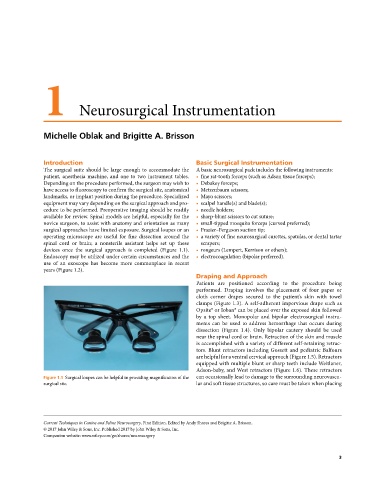
surgical approaches have limited exposure. Surgical loupes or an • Frazier–Ferguson suction tip;
operating microscope are useful for fine dissection around the • a variety of fine neurosurgical curettes, spatulas, or dental tartar
spinal cord or brain; a nonsterile assistant helps set up these scrapers;
devices once the surgical approach is completed (Figure 1.1). • rongeurs (Lempert, Kerrison or others);
Endoscopy may be utilized under certain circumstances and the • electrocoagulation (bipolar preferred).
use of an exoscope has become more commonplace in recent
years (Figure 1.2).
Draping and Approach
Patients are positioned according to the procedure being
performed. Draping involves the placement of four paper or
cloth corner drapes secured to the patient’s skin with towel
clamps (Figure 1.3). A self‐adherent impervious drape such as
Opsite® or Ioban® can be placed over the exposed skin followed
by a top sheet. Monopolar and bipolar electrosurgical instru-
ments can be used to address hemorrhage that occurs during
dissection (Figure 1.4). Only bipolar cautery should be used
near the spinal cord or brain. Retraction of the skin and muscle
is accomplished with a variety of different self‐retaining retrac-
tors. Blunt retractors including Gossett and pediatric Balfours
are helpful for a ventral cervical approach (Figure 1.5). Retractors
equipped with multiple blunt or sharp teeth include Weitlaner,
Adson‐baby, and West retractors (Figure 1.6). These retractors
Figure 1.1 Surgical loupes can be helpful in providing magnification of the can occasionally lead to damage to the surrounding neurovascu-
surgical site. lar and soft tissue structures, so care must be taken when placing
Current Techniques in Canine and Feline Neurosurgery, First Edition. Edited by Andy Shores and Brigitte A. Brisson.
© 2017 John Wiley & Sons, Inc. Published 2017 by John Wiley & Sons, Inc.
Companion website: www.wiley.com/go/shores/neurosurgery
3