
Page 16 - Zoo Animal Learning and Training
P. 16
Chapter 1: Neurosurgical Instrumentation 7
A
B
Figure 1.13 Hemostatics commonly used in neurosurgery: (top) bone wax;
(bottom left) gelatin sponge; (bottom right) cellulose surgical spears.
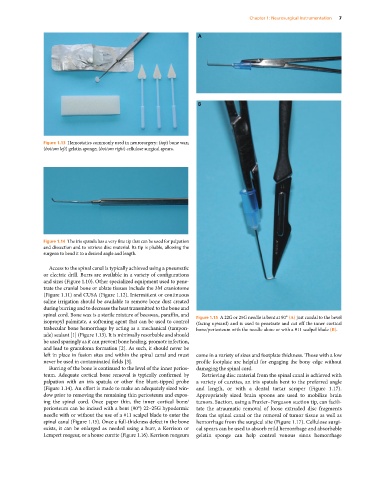
Figure 1.14 The iris spatula has a very fine tip that can be used for palpation
and dissection and to retrieve disc material. Its tip is pliable, allowing the
surgeon to bend it to a desired angle and length.
Access to the spinal canal is typically achieved using a pneumatic
or electric drill. Burrs are available in a variety of configurations
and sizes (Figure 1.10). Other specialized equipment used to pene-
trate the cranial bone or ablate tissues include the 3M craniotome
(Figure 1.11) and CUSA (Figure 1.12). Intermittent or continuous
saline irrigation should be available to remove bone dust created
during burring and to decrease the heat transmitted to the bone and
spinal cord. Bone wax is a sterile mixture of beeswax, paraffin, and Figure 1.15 A 22G or 25G needle is bent at 90° (A) just caudal to the bevel
isopropyl palmitate, a softening agent that can be used to control (facing upward) and is used to penetrate and cut off the inner cortical
trabecular bone hemorrhage by acting as a mechanical (tampon- bone/periosteum with the needle alone or with a #11 scalpel blade (B).
ade) sealant [1] (Figure 1.13). It is minimally resorbable and should
be used sparingly as it can prevent bone healing, promote infection,
and lead to granuloma formation [2]. As such, it should never be
left in place in fusion sites and within the spinal canal and must come in a variety of sizes and footplate thickness. Those with a low
never be used in contaminated fields [3]. profile footplate are helpful for engaging the bony edge without
Burring of the bone is continued to the level of the inner perios- damaging the spinal cord.
teum. Adequate cortical bone removal is typically confirmed by Retrieving disc material from the spinal canal is achieved with
palpation with an iris spatula or other fine blunt‐tipped probe a variety of curettes, an iris spatula bent to the preferred angle
(Figure 1.14). An effort is made to make an adequately sized win- and length, or with a dental tartar scraper (Figure 1.17).
dow prior to removing the remaining thin periosteum and expos- Appropriately sized brain spoons are used to mobilize brain
ing the spinal cord. Once paper thin, the inner cortical bone/ tumors. Suction, using a Frazier–Ferguson suction tip, can facili-
periosteum can be incised with a bent (90°) 22–25G hypodermic tate the atraumatic removal of loose extruded disc fragments
needle with or without the use of a #11 scalpel blade to enter the from the spinal canal or the removal of tumor tissue as well as
spinal canal (Figure 1.15). Once a full‐thickness defect in the bone hemorrhage from the surgical site (Figure 1.17). Cellulose surgi-
exists, it can be enlarged as needed using a burr, a Kerrison or cal spears can be used to absorb mild hemorrhage and absorbable
Lempert rongeur, or a house curette (Figure 1.16). Kerrison rongeurs gelatin sponge can help control venous sinus hemorrhage