
Page 152 - Zoo Animal Learning and Training
P. 152
Chapter 16: Dorsal Cervical Decompression (Laminectomy/Hemilaminectomy and Laminotomy) 153
a rich plexus of vascular structures surrounding the regions of the switches to burrs of smaller diameter as one advances more deeply.
articular facets, and brisk hemorrhage could ensue. Bipolar electro- Smaller burrs also facilitate undercutting of the inner cortical wall
cautery on appropriately sized bayonet forceps is helpful in control- and cancellous bone of the articular facets in dogs with hypertro-
ling bleeding vessels and oozing that occurs throughout the phy and bony stenosis of the vertebral canal, as occurs in young,
dissection down to the vertebral column. Tamponade can also be giant‐breed dogs with the osseous‐associated form of wobbler syn-
achieved by placement of moistened surgical sponges or laparot- drome. Surgeons vary in their preference to the use of irrigation
omy sponges (in larger patients) between self‐retaining retractors during high‐speed drilling. Continuous lavage has the advantage
and dissected muscles. of preventing thermal injury to the bone and adjacent tissues.
Performance of the laminectomy procedure itself and subsequent However, it can become very hard to visualize the burr-bone inter-
manipulations within the vertebral canal are enhanced by the use of face when continuously under liquid. I prefer to lavage intermit-
modest magnification (loupes) and possibly as well by supplement- tently, especially if operating without assistance. In either case, it is
ing overhead surgical lights with a head‐mounted illumination sys- vital to have suction at hand and a selection of various size tips
tem. Identification of the desired vertebra or vertebrae is based on (usually Frazier tips) in order to clear the surgical field of blood,
palpation or visualization of the spinous process of the axis crani- bone dust, and lavage fluid. The lateral extent of the laminectomy
ally, the markedly taller spinous process of T1, and then palpating is limited to just the medial‐most aspect of each articular facet.
or visualizing the much more truncated spinous processes of C3 This avoids causing excessive mechanical disruption, and also
through C7. reduces the risk for encountering the vertebral artery and its
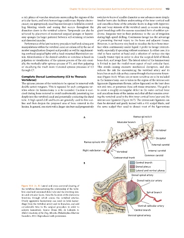
branches on each side as they course through the transverse foram-
Complete Dorsal Laminectomy (C3 to Thoracic inae (Figure 16.6). When two or more vertebrae are to be included
Vertebrae) in the laminectomy, care is taken in the region of the interarcuate
The spinous process of the vertebra to be opened is removed with ligaments (ligamentum flavum; yellow ligament) so the burr does
double‐action rongeurs. This is repeated for each contiguous ver- not sink into, or penetrate these soft tissue structures. The goal is
tebra where the laminectomy is to be extended. Caution is exer- to create a roughly rectangular defect in the outer cortical bone
cised during bone removal to avoid inadvertently penetrating too and cancellous bone of the lamina such that all that remains cover-
deeply into the vertebral canal and potentially contacting the dural ing the vertebral canal is the thin inner cortical bone layer and the
tube. A high‐speed drill and burrs are generally used to first out- interarcuate ligament (Figure 16.7). The interarcuate ligament can
line and then deepen the proposed area of bone removal in the then be elevated and gently incised with a #11 scalpel blade, and
lamina. In general, one starts with a larger‐size burr and progressively the same scalpel then used to dissect most of the ligamentous
A Ramus spinalis II
Muscular branch
Vertebral
Occipital
Ramus spinalis VIII
External carotid
Right subclavian Internal carotid
Vertebral
Common carotid
Costocervical trunk
B Central branch
Dorsal plexus
Lateral and ventral plexus
Dorsal spinal artery
Dorsal radicular artery
Figure 16.6 (A, B) Lateral and cross‐sectional drawing of
the vertebrae demonstrating the relationship of the verte-
bral canal and contained dural tube and the overlying lam- Spinal branch
ina and articular facets. Note the location of the transverse
foramina through which course the vertebral arteries.
Overly aggressive facetectomy can result in brisk hemor-
rhage from the vertebral artery and its branches, and add
considerable time to the surgical procedure in order to Ventral radicular artery
achieve hemostasis. Source: Evans HE, de Lahunta A. Central branch
Miller’s Anatomy of the Dog, 4th edn. Philadelphia: Elsevier
Saunders, 2013. Reproduced with permission. Ventral spinal artery