
Page 153 - Zoo Animal Learning and Training
P. 153
154 Section III: Spinal Procedures
A B
C
D
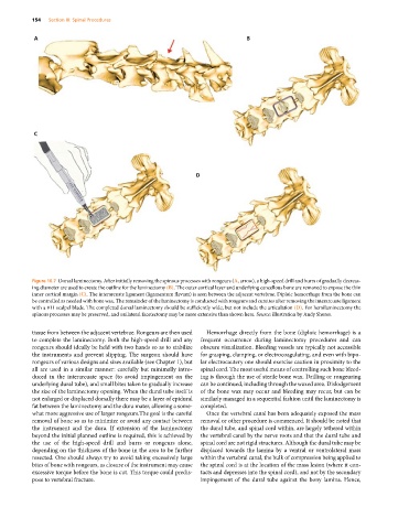
Figure 16.7 Dorsal laminectomy. After initially removing the spinous processes with rongeurs (A, arrow), a high‐speed drill and burrs of gradually decreas-
ing diameter are used to create the outline for the laminectomy (B). The outer cortical layer and underlying cancellous bone are removed to expose the thin
inner cortical margin (C). The interarcuate ligament (ligamentum flavum) is seen between the adjacent vertebrae. Diploic hemorrhage from the bone can
be controlled as needed with bone wax. The remainder of the laminectomy is conducted with rongeurs and curettes after removing the interarcuate ligament
with a #11 scalpel blade. The completed dorsal laminectomy should be sufficiently wide, but not include the articulation (D). For hemilaminectomy the
spinous processes may be preserved, and unilateral facetectomy may be more extensive than shown here. Source: illustration by Andy Shores.
tissue from between the adjacent vertebrae. Rongeurs are then used Hemorrhage directly from the bone (diploic hemorrhage) is a
to complete the laminectomy. Both the high‐speed drill and any frequent occurrence during laminectomy procedures and can
rongeurs should ideally be held with two hands so as to stabilize obscure visualization. Bleeding vessels are typically not accessible
the instruments and prevent slipping. The surgeon should have for grasping, clamping, or electrocoagulating, and even with bipo-
rongeurs of various designs and sizes available (see Chapter 1), but lar electrocautery one should exercise caution in proximity to the
all are used in a similar manner: carefully but minimally intro- spinal cord. The most useful means of controlling such bone bleed-
duced in the interarcuate space (to avoid impingement on the ing is through the use of sterile bone wax. Drilling or rongeuring
underlying dural tube), and small bites taken to gradually increase can be continued, including through the waxed area. Dislodgement
the size of the laminectomy opening. When the dural tube itself is of the bone wax may occur and bleeding may recur, but can be
not enlarged or displaced dorsally there may be a layer of epidural similarly managed in a sequential fashion until the laminectomy is
fat between the laminectomy and the dura mater, allowing a some- completed.
what more aggressive use of larger rongeurs.The goal is the careful Once the vertebral canal has been adequately exposed the mass
removal of bone so as to minimize or avoid any contact between removal or other procedure is commenced. It should be noted that
the instrument and the dura. If extension of the laminectomy the dural tube, and spinal cord within, are largely tethered within
beyond the initial planned outline is required, this is achieved by the vertebral canal by the nerve roots and that the dural tube and
the use of the high‐speed drill and burrs or rongeurs alone, spinal cord are not rigid structures. Although the dural tube may be
depending on the thickness of the bone in the area to be further displaced towards the lamina by a ventral or ventrolateral mass
resected. One should always try to avoid taking excessively large within the vertebral canal, the bulk of compression being applied to
bites of bone with rongeurs, as closure of the instrument may cause the spinal cord is at the location of the mass lesion (where it con-
excessive torque before the bone is cut. This torque could predis- tacts and depresses into the spinal cord), and not by the secondary
pose to vertebral fracture. impingement of the dural tube against the bony lamina. Hence,